ISSN: 0970-938X (Print) | 0976-1683 (Electronic)
Biomedical Research
An International Journal of Medical Sciences
- Biomedical Research (2013) Volume 24, Issue 1
A case report of coeliacomesenteric trunk
Department of Anatomy, Meram Medical Faculty, Necmettin Erbakan University, 42080 Meram, Konya, TURKEY
- *Corresponding Author:
- Mehmet Tugrul Yılmaz
Department of Anatomy, Meram Medical Faculty
Necmettin Erbakan University
42080 Meram, Konya, Turkey
Tel: +90 332
Accepted date: November 03 2012
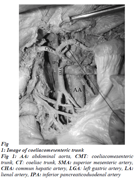
The coeliac trunk and superior mesenteric artery are the anterior visceral branches of the abdominal aorta. The anatomical variations of these arteries are due to development of the anterior splanchnic arteries. A case of coeliacomesenteric trunk was reported in this study which was observed in a 72-year-old male cadaver during the routine dissection. This trunk with a diameter of 13.98 mm is originated from the anterolateral wall of the abdominal aorta, 76.89mm below the aortic hiatus. After a 13.42 mm course, the trunk divided into coeliac trunk with a diameter of 7.09 mm and a superior mesenteric artery with the diameter of 5.25 mm. The coeliac trunk divided into splenic (6.85 mm diameter), common hepatic (5.31 mm diameter) and left gastric arteries (3.28 mm diameter). The superior mesenteric artery has inferior pancreaticoduodenal artery (3.20 mm diameter) arised from it as its first branch. Knowledge of variations of the coeliac trunk is important for both abdominal surgical approaches and radiological assessments.
Keywords
coeliacomesenteric trunk, variation, superior mesenteric artery, coeliac trunk
Introductıon
The coeliac trunk is the first branch of the abdominal aorta which originates from its anterior surface at the level of the 12th thoracal vertebra just below the aortic hiatus. It is a short and thick common root. It divides into three branches as left gastric, splenic and common hepatic arteries. It has been first described by Haller. Therefore, it is also known as “Tripus Haller”. The second branch that originated from the anterior surface of the abdominal aorta is the superior mesenteric artery and it generally divides from the abdominal aorta from 1.60 cm below the truncus coeliacus [1-6]. Several variations related to the localization of origins of coeliac trunk and superior mesenteric artery have been reported. One of these variations is that both arteries can originate from the abdominal aorta with a common trunk and therefore it is designated as the coeliacomesenteric trunk (CMT) [7-9].
Case report
In the 72-year-old male cadaver, the CMT was observed 76.89 mm below the diaphragma during the educational dissections. The CMT had a diameter of 13.98 mm and originated from the anterior surface of the abdominal aorta just below the aortic hiatus. The length of the CMT was found to be 13.42 mm and it divided into two branches as the coeliac trunk and superior mesenteric artery. The coeliac trunk with its own diameter of 7.09 mm divided into three branches as the common hepatic, left gastric, and lienal arteries. The diameters of these three branches were 5.3 mm, 3.28 mm, and 6.85 mm, respectively. In addition , the root diameter of the superior mesenteric and the diameter of the inferior pancreaticoduodenal arteries were 5.25 mm and 3.20 mm respectively with the latter branch serving as the first branch of the former (Figure 1).
Dıscussıon
The knowledge of the arterial variations are important in terms of the development and application of the new invasive surgical techniques, organ transplantations, and the diagnosis, prevention, and treatment of some metastatic tumours [1,5,6,10,11]. The incidence of the normal trifurcated structure of the coeliac trunk which is the origin of the common hepatic, left gastric, and lienal arteries is 86% while those of the bifurcated structure and the absent coeliac trunk are 12% and 2%, respectively. Beside this branching, bifurcated structure of CT (12%) and nonexistence of CT (2%) shouldn’t be ruled out [1-4,6]. On the other side, the variations of the superior mesenteric artery which originate from the anterior surface of the abdominal aorta are rare. However, the variations of the superior mesenteric artery associated with the coeliac trunk and its branches were especially mentioned in the literature [2,9-12]. Demirtas et al. [10] have reported a case that coeliac trunk has formed two trunks, such as hepatogastric and hepatosplenic trunks, without a trifurcation structure. In the Japanase cadaver of a 79-year-old female, Saga et al. [11] have found the hepatomesenteric trunk that originated from the abdominal aorta with a common root. The case of the absent of the coeliac trunk was reported by Yamaki et al. [12]. In such case, the common hepatic, left gastric, and lienal arteries have arised from the abdominal aorta by their independent origins. Emirzeoğlu et al. [13] have observed the left gastric and lienal arteries separately arised from the abdominal aorta whereas the common hepatic and superior mesenteric arteries originated from the abdominal aorta with a common root in a 42-year-old patient with pre-diagnosis of mesenteric ischemia. The cases of the CMT indicate no specific symptoms clinically but they may be generally realized during retrospective screenings, surgical procedures or dissection studies. Ailawadi et al. [7] determined the CMT in 18 (14 male and 4 female) patients during retrospective screening. Four of the cases had applied due to the circulatory system disorders, whereas the remaining fourteen patients had applied because of nonvascular symptoms of gastrointestinal system, chemotherapy, lower extremity pain or they have been diagnosed accidentally. Lezzi et al. [2] have reported the coeliac trunk with normal anatomical structure in 72.1% of the cases, hepatogastric trunk in 5%, splenogastric trunk in 3.6%, hepatosplenic trunk in 2.7%, and hepatosplenomesenteric trunk 0.4% on 524 MDCT images. Besides, it has been detected that the branches of the coeliac trunk originate separately from the abdominal aorta or the superior mesenteric artery without forming a trifurcation structure.
CMT originated from anterior surface or anterolateral side of the abdominal aorta has been reported in several cadaver studies [6,8,9,14]. In this study, the CMT have been arised from the anterior surface of the abdominal aorta. Pennington et al. [8] have reported that the origination diameters of the coeliac trunk and the superior mesenteric artery as 0.76 cm (0.54-1.08 cm) and 0.91 cm (6.2-1,21 cm), respectively. In our case, the origination diameters of the coeliac trunk and the superior mesenteric artery were 0.70cm and 0.52cm, respectively.
Conclusıon
In the cases of CMT, the potential acute or chronic mesenteric ischemia may be observed although no clinical symptoms were generally observed. The ischemia may be detected in the proximal end of intestinal system because of the insufficient arterial blood circulation between the coeliac trunk and the superior mesenteric artery. It should be kept in mind that such variations can be seen during operations and it must be on the lookout for in this group of patients carefully and meticulously.
References
- Ferrari R, Cecco CN, Iafrate F, Paolantonio P, Rengo M, Laghi A. Anatomical variations of the coeliac trunk and the mesenteric arteries evaluated with 64-row ct angiography. Radiol Med 2007; 112: 988-998.
- Lezzi R, Cotroneo RA, Giancristofaro D, Santoro M, Storto ML. Multidetector-row ct angiographic imaging of the celiac trunk: anatomy and normal variants. Surg Radiol Anat, 2008; 30: 303-310.
- Vandamme JPJ, Bonte J. The branches of the celiac trunk. Acta Anat 1985; 122: 110-114.
- Warwick R, Williams LP. Gray’s Anatomy, Elsevier Churchill Livingstone. Thirty ninth Edition, 2005; 1118-1120.
- Yahel J, Arensburg B. The topographic relationships of the unpairedvisceral branches of the aorta. Clin Anat 1998; 11: 304-09.
- Yüksel M, Sargon M. A variation of a coeliac trunk. Okajimas Folia. Anat Jpn 1992; 69: 173-176.
- Ailawadi G, Robert AC, James CS, Jonathan LE, David MW, Lisa MC, Peter KH, Gilbert RU. Common celiacomesenteric trunk: Aneurysmal and occlusive disease. Journal of Vascular Surgery 2004; 40(5): 1040-1043.
- Çicekcibasi AE, Uysal ĐĐ, Seker M, Tuncer I, Büyükmumcu M, Salbacak A. A rare variation of the coeliac trunk. Ann Anat 1997; 187: 387-391.
- Yi SQ, Terayama H, Naito M, Hayashi S, Moriyama H, Tsuchida A, Itoh M. A common celiacomesenteric trunk, and a brief review of the literature. Ann Anat 2007; 189: 482-488.
- Demirtas K, Gulekon N, Kurkcuoglu A, Yıldırım A, Gozil R. Rare variation of the celiac trunk and related reiew. Saudi Med J 2005; 26 (11): 1809-811.
- Saga T, Hirao T, Kitashima S, Watanabe K, Nohno M, Arakı Y, Kobayashi S, Yamaki K. An anomalous case of the left gastric artery, the splenic artery and hepatomesenteric trunk independently arising from the abdominal aorta. Kurume Medical Journal 2005; 52: 49-52.
- Yamaki K, Tanaka N, Matsushima T, Miyazaki K, Yoshizuka M. A rare case of absence of the celiac trunk: the left gastric, the splenic, the common hepatic and the superior mesenteric arteries arising independtly from the abdominal aorta. Ann Anat 1995; 177: 97-100.
- Emirzeoğlu M, Dinç H, Arslan MK, Uluutku MH, Yeğinoğlu G. A case in which common hepatic artery and superior mesenteric artery from a trunk together and no celiac trunk is formed. Morfoloji Dergisi 1997; 5(1-2): 13-14.
- Çavdar S, Sehirli U, Pekin B. Celiacomesenteric trunk. Clin Anat 10: 231-234.
- Pennington N, Soames RW. The anterior visceral branches of the abdominal aorta and their relationship to the renal arteries. Surg Radiol Anat, 2005; 27: 395-403.
