ISSN: 0970-938X (Print) | 0976-1683 (Electronic)
Biomedical Research
An International Journal of Medical Sciences
Case Report - Biomedical Research (2022) Volume 33, Issue 1
A giant cutaneous squamous cell carcinoma in adolescent: A case report and a brief literature review.
Khadija Darif*, Samia Arifi, Reda-El-Hazzaz, Oumaima Siyouri, Khadija Hinaj, Lamiae Amaadour, Karima Oualla, Zineb Benbrahim, Fz El M'rabet, Nawfel Mellas
Department of Medical Oncology, Hassan II University Hospital Center, Faculty of Medicine and Pharmacy of FEZ, Sidi Mohamed BenAbdellah University, Fez, Morocco
- Corresponding Author:
- Khadija Darif
Department of Medical Oncology
Hassan II University Hospital Center
Fez, Morocco
Accepted date: January 03, 2022
Cutaneous squamous cell carcinoma is very rare skin pathology in children and adolescents, most of the cases reported in the literature are small lesions that occur under predisposing conditions. Here we report a rare case of a teenager with a huge tumor without predisposing factors. The patient presented at Hassan II University Hospital with a huge mass with active bleeding. The imaging showed a locally advanced mass invading the scapular humeral joint, with lung metastasis. Histology revealed a cutaneous epidermoid carcinoma. Hemostatic radiation therapy has been performed followed by a systemic chemotherapy based on a platine regim.
The aim of this paper is to describe the clinical and radiological particularities of a very challenging case of a young patient with a skin squmous cell carcinoma.
Keywords
Skin Squamous, Cell Carcinoma, Giant Tumor, Adolescent.
Introduction
Skin Squamous Cell Carcinoma (SCC) is a very rare skin cancer in children and adolescents [1]. Most cases occur in the context of underlying predisposing conditions such as UV light, ionizing radiation, predisposing genodermatoses, and Long-term immunosuppression.
In this article we report a rare case with an atypical presentation of squamous cell carcinoma, treated in the oncology department of the Hassan II University Hospital of Fez.
Case report
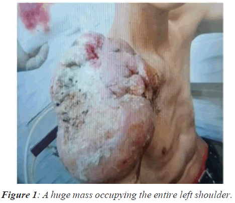
We report a rare case of cutaneous squamous cell carcinoma in 18 years old boy, with no pathological history. The teenager presented a nodule of the left shoulder which was devloping for 12 months in line with a weight loss, without any traumatic incident or predisposing factor. The lesion progressed rapidly; it changed from a small subcutaneous nodule of a few millimeters at the start to a huge mass of 30 cm occupying the entire left shoulder (Figure 1), the patient also presented several bleeding episodes which inited the patient to go to the emergencies suffering from deep anemia; It was his first consultation which came at a very progressive stage.
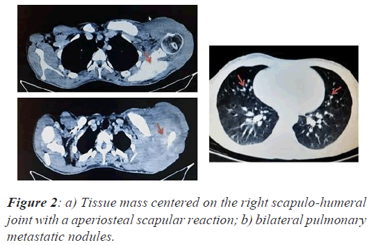
A thoraco abdomino pelvic CT scan was also performed revealing a Tissue mass centered on the right scapulo- humeral joint extended along the homolateral clavicule measuring 15 x 19 x 17 cm (Figure 2A).This mass is responsible for a periosteal scapular reaction and a diffuse bone condensation of the clavicule and seems to invade the thoarcic wall with regard to 2,3 and 4th rib ; with bilateral pulmonary metastasis (Figure 2B).
A biopsy of the tumor has been performed, histology revealed a malignant small to medium sized round cells proliferation ; Immunohistochemistry showed an intense expression of cytokeratins CK5, CK6 and the P63 protein .However, TTF1, CD30, CD20, CD117 staining were. Pathologist concluded to poorly differentiate epidermoid carcinoma.
Clinical and radiological workup including head and neck examination and chest scanning, does not reveal a primary tumour, A second anatomopathological review has been requested and confirmed the diagnosis of an epidermoid carcinoma. A primary skin squamous carcinoma with lung metastasis has been retained.
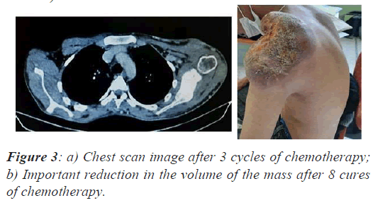
The patient underwent hemostatic radiation therapy 10gy (5gy*2) then he was put on palliative chemotherapy with carboplatin paclitaxel regim (carboplatine AUC5, paclitaxel 175 mg / m2 ever 21 days). After 3 months the patient has improved clinically with a decrease in the volume of the tumor mass, reduction in pain and cessation of bleeding, Radiological evaluation objectived a partial response according to the Response Evaluation Criteria In Solid Tumours (RECIST) version 1.1 , therefore chemotherapy was continued, after 8 cycles, the clinical and partial radiological response is maintained (Figure 3A-3B).
Discussion
Nonmelanoma skin (SCC, BCC (basal cell carcinoma)) cancers are very rare in children and adolescents. There are few scientific data regarding this pathology and its management .SCC is a malignant tumor of squamous cells of the epidermis characterized by rapid progression, as well as greater invasive and metastatic ability compared to BCC. SCC occurs more often on the sun-exposed areas, others sites are very uncommon for this type of carcinoma [8].
In a report of 7,814 cases of primary skin cancers in individuals younger than 30 years who were recorded by the Surveillance, Epidemiology, and End Results database from 2000 to 2008, carcinomas accounted for 0.008% of all cases [2,5]. In a study from the north of England, only 6 SCCs of the skin were seen in 28 years [7].
Most of the cases reported in the literature had predisposing skin conditions: in one serie of 28 patients, approximately one-half of patients had predisposing conditions such as nevoid basal cell carcinoma syndrom and one-half of patients were exposed to iatrogenic conditions such as prolonged immunosuppression or radiation [3,5].
Another serie of tumor skin in children between 1992 and 2006 found three patients with cutaneous SCC and all of these patients had predisposing skin conditions namely xeroderma pigmentosa, sebaceous naevus and dystrophic epidermolysis bullosa [4].
One case of cutaneous squamous cell carcinoma in a child with no history of predisposing conditions, repported in 7-year-old girl ; the patient had a small lesion and she benefited from excision with negative margins [5].
Regarding clinical presentation, SCC are usually reddened lesions with varying degrees of scaling or crusting, and they have an appearance similar to eczema, infections, trauma, or psoriasis [6].
Treatment of Childhood SCC of the Skin is mainly surgical excision or Mohs micrographic surgery, [6] when it is generally a small lesion; in our case the tumor was very locally advanced therefore not resectable very little data concerning the systematic treatment in the literature.
The characteristics that make our case a rarity are the absence of any underlying skin pathology with an unusual clinical presentation of cutaneous squamous carcinoma as well as therapid and aggressiveevolution at thelocoregional level and at a distance, but despite the aggressiveness and the extension of the disease chemotherapy was effective leading to an objective response with the improvement of the patient's quality of life for one year until now.
Conclusion
The delay in diagnosis as well as the rapid and aggressive development of the disease made the treatment of the patient very complicated, in spite of the rarity of this pathology, it is still necessary to biopsy any suspicious skin lesion in adolescents spacialy in patients with a history of immunosuppression, tumours, radiotherapy, or genetic predisposition to malignancies.
Abbreviations
SCC: Skin squamous Cell Carcinoma; RECIST: Response Evaluation Criteria In Solid Tumours; BCC: Basal Cell Carcinoma
Declarations
Written informed consent was obtained from the patient for publication of this case report and any accompanying images. A copy of the written consent is available for review by the Editor-in-Chief of this journal.
Competing Interests
I declare no link of interest
Availability of Supporting Data
All data is available
Funding
I do not declare any funding
References
- Henning Hamm, Peter H Hoger. Skin Tumors in Childhood. Dtsch Arztebl Int 2011; 108: 347-353.
[Crossref] [Google Scholar] [PubMed]
- Senerchia AA, Ribeiro KB, Rodriguez-Galindo C. Trends in incidence of primary cutaneous malignancies in children, adolescents, and young adults: A population-based study. Pediatr Blood Cancer 2014; 61: 211-216.
[Crossref] [Google Scholar] [PubMed]
- Khosravi H, Schmidt B, Huang JT. Characteristics and outcomes of Nonmelanoma Skin Cancer (NMSC) in children and young adults. J Am Acad Dermatol 2015; 73: 785-790.
[Crossref] [Google Scholar] [PubMed]
- Chow CW, Tabrizi SN, Tiedmenn K. Squamous cell carci- nomas in children and young adults: A new wave of rare tumour. J Pediatr Surg 2007; 42: 2035-2039.
[Crossref] [Google Scholar ] [PubMed]
- Kotwal A, Watt D. Cutaneous squamous cell carcinoma in a child. J Plast Reconstr Aesthet Surg 2009; 62: e194-e195.
[Crossref] [Google Scholar] [PubMed]
- PDQ Pediatric Treatment Editorial Board. Childhood Basal Cell Carcinoma and Squamous Cell Carcinoma of the Skin Treatment (PDQ®): Patient Version. 2020.
- Pearce MS, Parker L, Cotterill SJ. Skin cancer in children and young adults: 28 years' experience from the Northern Region Young Person's Malignant Disease Registry, UK. Melanoma Res 2003; 13: 421-426.
[Crossref] [Google Scholar] [PubMed]
- Melo Neto B, Oliveira GP, Vieira SC, Leal LR, Melo Jr JAC, Vie ira CF. Epidermoid carcinoma of the skin mimicking breast cancer. An Bras Dermatol. 2013; 88: 250-252.
[Crossref] [Google Scholar] [PubMed]


