ISSN: 0970-938X (Print) | 0976-1683 (Electronic)
Biomedical Research
An International Journal of Medical Sciences
- Biomedical Research (2016) Volume 27, Issue 2
Alterations in the anterior segment of the eye and in central corneal thickness in the early periods following blunt eye trauma.
| Kursat Atalay1*, Havva Erdogan1, Ahmet Kirgiz1, Metin Mert1, Ceren Gurez1, Muhittin Taskapili2 1Bagcılar Education and Research Hospital, Eye Clinic, 34200, Istanbul 2Prof.Dr. Resat Belger Beyoglu Education and Research Hospital, Istanbul |
| Corresponding Author: Kursat Atalay, Bagcilar Education and Research Hospital Turkey |
| Accepted: January 30, 2016 |
Aim: To use ultrasonographic pachymetry and Anterior Segment Optic Coherence Tomography (ASOCT) to determine changes in anterior segment structures and Central Corneal Thickness (CCT) in patients within 48 hours of blunt eye trauma. Our overall goal was to identify the effects of CCT alterations on blurred vision during the acute periods of blunt eye trauma.
Method: Anterior segment findings were recorded for all patients. CCT measurements of 43 traumatized eyes were compared for 43 traumatized eyes and 41 non traumatized eyes. CCT was measured with a USG pachymeter and AS-OCT.
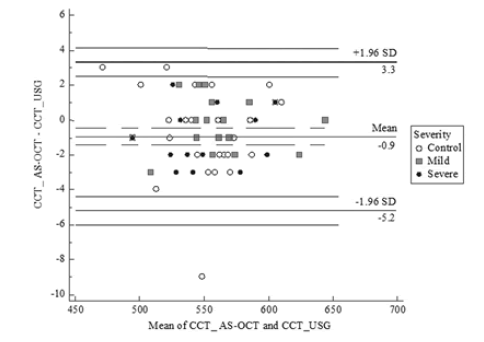
Results: The mean best corrected visual acuities differed significantly between the control group and patients sub-grouped as mildly and severely injured (p=0.006).The mean IOP was higher for the mildly traumatized group than for the control group,butthe difference was not statistically significant (p=0.53).The mean IOP increased in the severely traumatized group when compared to the control and mildly traumatized groups (p=0.001, p=0.035; respectively).USG pachymetry and AS-OCT measurements were not significantly different among the three groups(p=0.43, p=0.38; respectively). Both measurement methods showed good correlation in the control, mildly traumatized, and severely traumatized groups (r=0.997, p=0.000; r=0.999, p=0.000; r=0.998, p=0.000; respectively). Bland-Altman analysis revealed a high level of agreement between USG pachymetry and AS-OCT in every sub-group.
Conclusion: We found no significant differences in the CCT values between healthy and traumatized eyes. Therefore, AS-OCT may be a good alternative protocol for examination of acutely traumatized eyes due to the decreased contamination risk.
Keywords |
||||||
| Trauma, Eye, Central corneal thickness, Optic coherence tomography | ||||||
Introduction |
||||||
| The cornea is a unique tissue due to its refractive and translucent properties. The corneal endothelium plays an essential role in preserving corneal transparency; however, the endothelial cells are arrested in the G1phase of the cell cycle and therefore do not regenerate. Any loss of cells is instead compensated by the expansion and spreading of the remaining cells [1]. The resulting reduction in the overall cell density of the endothelium causes disturbances in the fluid balance, which then results in stromal swelling and ultimatelyin transparency loss and impaired vision. Thus, the prevention, early detection, and immediate treatment of corneal endothelium dysfunction are extremely important for sustained vision. | ||||||
| The corneal thickness and cell density can be used asindirect measures of endothelial function, and they reflect clinically important and directeffects of toxins on the endothelium [2]. Corneal thickness is determined by pachymetric measurements using different types of pachymetry protocols, such as USGP and AS-OCT. Ocular trauma, although preventable in the majority of cases, is one of the main causes of blindness and disability in developing countries [3]. Balaghafari et al., reported that 12.4% of 178 cases of ocular trauma in their study were due to blunt objects [4]. Blunt eye trauma can lead to severe complications, including impaired vision, contusions involving the anterior segment, and lens injuries, such as contusion cataracts and partial or total dislocation [5,6]. | ||||||
| The aim of the presentstudy was to use USGP and AS-OCT to determine the changes in the anterior segment of the eye and in Central Corneal Thickness (CCT) in patients within 48 hours of blunt eye trauma. Our goal was to identify the effects of CCT alterations on blurred vision during the acute periods of blunt eye trauma. | ||||||
Material and Methods |
||||||
| This prospective cross sectional study was designed to evaluate the patients who were admitted between January 2014 and June 2015 to Bagcilar Education and Research Hospital with the complaint of blunt eye trauma within 48 hours of the trauma. Informed consent was obtained from all participants and permission was received from the local ethics committee in compliance with the Helsinki declaration rules prior to the study. Patients who had a previous diagnosis of glaucoma, corneal degenerative diseases, previous ocular surgeries, contact lens use, or corneal scarring were excluded. The nontraumatized eyes of the same patients were used as the control group. The traumatized eyes of the patients were further divided into severely traumatized or mildly traumatized subgroups. Patients with traumatic eye findings without lid hematomas were grouped as mildly traumatized. Patients presenting with an eyelid hematoma and any additional pathologic findings were classified as severely traumatized. | ||||||
| Routine ophthalmologic examinations, including best corrected visual acuity (BCVA) tested with a Snellen chart, intraocular pressure measurements, and biomicroscopic and fundoscopic examinations were performed on all patients. Traumatic eye findings were noted, including eye lid hematoma, edema, ecchymosis, subconjunctival hemorrhage, Vossius ring, anterior chamber cells, angle recession, hyphema, and cataract formation. Inflammatory cells in the anterior chamber were graded as 0, +1, +2, or +3 by slit lamp biomicroscopy. | ||||||
| A total of 42 patients were included in this study. Examination of one patient showed a corneal epithelial defect, anterior displacement of the lens, cataract formation, and corneal lenticular touch. He could not fixate, so we were unable to measure the CCT with AS-OCT. We evaluated the CCT of all patients, except this particular patient. One patient out of 42 was injured in both eyes; therefore, both eyes were included as traumatized eyes, so that a total of 43 eyes were examined in the study group. Additional diagnostic tests, such as orbital tomography, were conducted when necessary. The Snellen measurements were converted to Log Mar for statistical purposes [7]. The central corneal thickness was measured by USGP and AS-OCT. An AccuPach (Accutome Ultrasound Inc.) device was used for USGP and the mean of three USGP measurements was recorded. A spectral domain OCT (Nidek Co. RS-3000) was used for the measurements of AS-OCT. The central corneal regions of the eyes were measured. All measurements were performed by the same examiner (K.A.). | ||||||
Statistical analysis |
||||||
| Data entry and analysis were done using the Statistical Package for Social Sciences SPSS version 16. The normality of the data was checked with a one-sample Kolmogorov-Simirnov test. When the data were normally distributed, a one way ANOVA test was used for comparison of the means. If the data were not normally distributed, the Kruskal Wallis and Mann Whitney-U tests were used. The Pearson correlation was used for correlation analysis of USGP and AS-OCT data. A Bland- Altman analysis was performed to determine the agreement between USGP and AS-OCT. A Chi-square test was used to analyze the differences in anterior chamber cell density. The analysis was considered to show a statistically significant association when the P-value was <0.05. | ||||||
Results |
||||||
| The demographic data of the study are displayed in Table 1. The mean BCVA for thecontrol group was 0.012 ± 0.051 (range: 0-0.3), and it was 0.055 ± 0.091 (range: 0-0.3) and 0.124 ± 0.23 (range: 0-1.0) for the mild and severe groups, respectively. The BCVA data were not normally distributed. The Kruskal Wallis analysis showed a significant difference between the three groups (p=0.006). The Mann Whitney U test showed a significant reduction in the BVCA of both the mildly and severely injured groups when compared to the control group (p=0.01; p=0.002, respectively). The difference between the mean BCVAs of the mildly and severely traumatized groups was not statistically significant (p=0.42). | ||||||
| The mean Intraocular Pressures (IOP) were 15.8 ± 2.8 mmHg (between11-25 mmHg) for the control group,16.7 ± 2.6 mmHg (between 11-23 mmHg) for the mildly injured group, and 19.2 ± 4.4 mmHg (between 12-30 mmHg) for the severely injured group. | ||||||
| Although the mean IOP of the mildly traumatized group was slightly higher than that of control group, the difference was not statistically significant (p=0.53). However, the mean IOP of the severely traumatized group was statistically higher than that of the control group or the mildly traumatized group (p=0.001, p=0.035; respectively). | ||||||
| Some of the anterior chamber findings were classified according to severity of the trauma and are listed in Table 2. The presence of inflammatory cells in the anterior chamber, as a result of traumatic iritis, was more prominent in the severely traumatized eyes. Noinflammatory cells were found in the nontraumatized eyes. Chi-square analysis of the anterior chamber inflammatory cell density showed a significant association with the severity of the trauma (p=0.012). | ||||||
| Table 3 shows the results of CCT measurements with USGP and AS-OCT. Comparison of the USGP measurements by oneway ANOVA analysis did not reveal any significant difference among the three groups (p=0.43). Similarly, comparison of the AS-OCT measurements of all three groups by one-way ANOVA analysis also did not reveal any statistically significant differences (p=0.38). | ||||||
| A strong correlation was found between the two CCT measurement methods for the control, mildly traumatized, and severely traumatized groups (r=0.997, p=0.000; r=0.999, p=0.000; r=0.998 p=0.000; respectively). The Bland-Altman analysis revealed a high level of agreement between USGP and AS-OCT in every sub-group (Figure 1). | ||||||
Discussion |
||||||
| In this study, we investigated the changes in CCT occurring within the first 48 hours following ablunt trauma. Our findings indicate that, although no statistically significant changes occurred in the CCT, the mean BCVA values were statistically significantly decreased after trauma. In this respect, the impaired or blurred vision occurring after blunt trauma would not appear to be associated with the alterations in the CCT in the early periods following a blunt trauma. | ||||||
| The available literature identifies several potential factors that may affect the CCT, including age, gender, ethnicity, diurnal variation, contact lens wear, and ocular surgery [8-11]. Some reports on CCT measurements have shown a decrease in the CCT with increased age, while others have reported no such relationship [12,13]. Our correlation analysis determined that age was not related with the CCT. Moreover, no statistically significant difference was noted between the groups in the present study in terms of age or gender; all participants shared the same ethnicity and had no previous history of contact lens wear or ocular surgery. | ||||||
| Blunt eye trauma can lead to severe complications, including loss of vision. Jasielska et al., who studied 15 patients with blunt ocular trauma and lens luxation to the vitreous, reported the mean IOP of patients as 20.6 mmHg at admission, before treatment, which is slightly higher than our findings [14]. They also determined the mean BCVA as 0.24 and 0.33, before and after surgical treatment, respectively. To the best of our knowledge, the direct effects of blunt eye trauma on the CCT have not been studied previously. Aribaba et al., analyzed 400 eyes of 200 patients and reported an increase in the mean baseline CCT 24 hours after cataract surgery. This was followed by a relative reduction in the mean CCT over a12 week period in both the operated and unoperated eyes [15]. They concluded that cataract surgery is a trauma that induces asystemic inflammatory response and inflammation mediators may play a role in the observedalterations in the CCT. | ||||||
| The CCT measurements can be conducted using several methods, such as USGP, the Scheimpflug technique, and OCT, and each technique has its own advantages and disadvantages [13]. The ultrasound-based technique is considered to be the gold standard, but it still has some drawbacks. First, it is a contact technique and can cause discomfort to patients; second, it does not enable investigators to obtain a corneal pachymetry map, nor can it identifythe thinnest point of the cornea. The Scheimpflug technique and OCT methods are non-contact techniques and reveal the iridocorneal angle [16]. The ASOCT technique, used in the present study, is based on the optic properties of light. It causes no discomfort to the patient, it is easy for clinicians to perform, and the AS-OCT pachymetry measurements are comparable with those obtained using other techniques [17]. We found a good correlation between our USGP and AS-OCT results. Since even healthy patients may experience ocular discomfort and contamination with USGP, AS-OCT may be a particularly good alternative to USGP for traumatized patients. | ||||||
| The reported CCT alterations can significantly affect intraocular pressure measurements obtained with the Goldmann applanation tonometer [18,19]. A correction for IOP, calculated from corneal thickness, has been defined previously [20,21]. Acute intraocular pressure increments due to blunt eye trauma can occur from mild traumatic iritishyphema to angle recession, and this increase in IOP may need treatment. Trauma-associated CCT alterations may become important when giving anti-glaucomatous medication to trauma patients and in their follow up. However, in the present study, we did not detect any alterations in either the CCT or the IOP within 48 hours after blunt trauma. Furthermore, we did not determine any correlation between the CCT and the IOP. | ||||||
| The present study hassome limitations that should be mentioned. The sample size was too small to make any generalizations. Second, we checked the effects of blunt trauma only during the first 48 hours after the trauma and we did not followup the patients. The CCT could possibly show more substantial alterations several days later after the trauma. However, we can conclusively say that changes in the BCVA values in the first 48 hours after a blunt trauma are not directly associated with the alterations in the CCT. | ||||||
| Significant diurnal fluctuations in the CCT have been reported in the literature in subjects without any ocular pathology. The CCT is defined as thickest in the morning, upon awakening, and it gradually thins as the day progresses; the greatest proportion of this variation occurs in the 3 hours after awakening [22,23]. Due to the study design, the CCT measurements were taken when the patients were admitted to the clinic, within 48 hours of the trauma, and therefore any circadian variations in the CCT were not taken into account. | ||||||
Conclusion |
||||||
| This prospective cross sectional study used USGP and the ASOCT to determine the changes in the CCT within 48 hours after blunt eye trauma. To the best of our knowledge, this is the first report in the literature to investigate the CCT using ASOCT within 48 hours of a blunt eye trauma. We did not detect any significant differences with respect to the CCT values between healthy and traumatized eyes. Larger studies are warranted to determine the effects of blunt eye trauma on the CCT, since defining the etiology will aid in managing blurred vision more appropriately during the acute periods following blunt trauma. | ||||||
Tables at a glance |
||||||
|
||||||
Figures at a glance |
||||||
|
||||||
References |
||||||
|
