ISSN: 0970-938X (Print) | 0976-1683 (Electronic)
Biomedical Research
An International Journal of Medical Sciences
Research Article - Biomedical Research (2020) Volume 31, Issue 5
Association of maternal age with cytogenetic variants of Down's Syndrome in North Indian population
Kanchan Bisht1, Rakesh K. Verma1*, Navneet Kumar1, Shakal N. Singh2, Baibhav Bhandari3
1Department of Anatomy, King George’s Medical University, Lucknow, Uttar Pradesh, India
2Department of Paediatrics, King George’s Medical University, Lucknow, Uttar Pradesh, India
3UCHC, N.K Road, Hazratganj, Lucknow, Uttar Pradesh, India
- Corresponding Author:
- Rakesh K. Verma
Department of Anatomy
King George’s Medical University
Lucknow
Uttar Pradesh
India
Accepted date: September 21, 2020
Background: Down’s syndrome is the most commonly encountered autosomal aneuploidy in the population involving chromosome 21. It is regarded as the most common genetic cause of intellectual disability in the world and is found to be associated with multiple risk factors. Objectives: The aim of our study was to observe the association of maternal age with cytogenetic variants of Down’s syndrome in North Indian population. Material and methods: The study was conducted in the cytogenetic laboratory of the Department of Anatomy, King George’s Medical University Uttar Pradesh, Lucknow. For the present study, peripheral blood samples were collected from 51 suspected patients of DS, of age group 0-10 years who were screened in Pediatrics Department, KGMU. Karyograms were prepared and evaluation was done. Results: Advanced maternal age was seen to be more closely associated with mosaic variant and least associated with translocation variant of Down’s syndrome.
Keywords
Down’s syndrome, Cytogenetic variants, Karyograms, Trisomy, Congenital.
Introduction
The syndrome got its name after a British physician, John Langdon Down who first recognized the syndrome in 1866. But its real cause was later understood in 1959, which led to the conclusion that an extra copy of chromosome 21 is responsible for this condition [1].
Down's syndrome (DS) is the most common genetic cause of mental retardation in the world. These patients usually have a characteristic phenotype like protruding tongue, microgenia, short stature [2], brachycephaly, flat facies, upward slanting palpebral fissures, epicanthus, low-set ears with abnormal folds, and simian crease [3]. This typical presentation of DS patients is often very helpful, making their clinical diagnosis relatively easy. Down’s syndrome has been linked with many systemic anomalies like mental retardation, cardiac anomalies, gastrointestinal anomalies, ophthalmological errors, hearing loss, skin problems and genitourinary abnormalities [2].
Various risk factors have been found to be associated with the occurrence of DS like advanced maternal age, consanguineous marriage, use of contraceptive pills, early induced abortion, socioeconomic conditions, cigarette smoking, alcohol intake and radiation exposure etc [4]. However, advanced maternal age is still considered as the principal risk factor for trisomy 21 [5]. If the mother is 35 years of age or older at the time of delivery, it is considered as advanced maternal age. [1] With late marriages being so common in present scenario, advanced maternal age is becoming an increasing unavoidable risk factor day by day, for giving birth to child with Down syndrome. Likelihood of having a DS baby rises from 1 in 1300 (in pregnant women under 25 years) to 1 in 300 (in pregnant women of 36 years) to 1 in 10 (in pregnant women of 49 years) [6]. Among older women available antral follicles are limited and ovary has to compromise in selecting a suboptimal or erroneous oocyte for ovulation. On the contrary, many studies report that majority of DS babies are born to young women of less than 30 years. Possible explanation of younger mothers having more children with DS could be usage of alcohol, tobacco, environmental toxins and drugs, improper sleep and diet [7].
The three main cytogenetic variants of Down’s syndrome are: Free trisomy 21, where an extra chromosome 21 is present in all the cells; Mosaic trisomy 21, in which two type of cell lineages are present, one is normal and another cell line with trisomy 21; Robertsonian translocation trisomy 21, where the long arm of chromosome 21 is attached to another chromosome (usually an acrosome) [8].
All the variants of DS do not present with similar features. One of the variants of DS, Mosaicism, shows milder phenotypes in comparison to free trisomy.
Materials and Methods
For the present study, peripheral blood samples were collected from 51 suspected patients of DS, of age group 0-10 years, screened in Department of Paediatrics, KGMU. Informed consent was taken from their parents/ guardians. While collecting samples, detailed personal history, family history and thorough clinical examination was done. The samples were cultured and karyograms were prepared in the Cytogenetic laboratory of the Department of Anatomy, King George’s Medical University U.P., Lucknow.
This descriptive cytogenetic study was ethically approved by the Ethical committee of King George’s Medical University U.P., Lucknow
Data was analysed using Statistical package for Social Science (SPSS) version 21.0. Data has been represented as frequencies and percentages and mean and standard deviation. Chi-square test has been used for the purpose of analysis. The confidence level of the study was kept at 95%, hence a “p” value less than 0.05 indicated a significant association.
Observation and Results
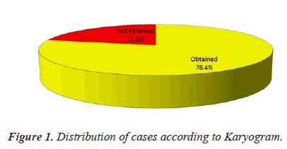
A total of 51 subjects were screened, however, karyogram could be obtained for only 40 (78.4%) subjects (Table 1 and Figure 1).
| SN | Outcome | Statistic |
|---|---|---|
| 1. | Obtained | 40 (78.4%) |
| 2. | Not obtained | 11 (21.6%) |
Table 1: Karyogram Obtained/Not obtained (n=51).
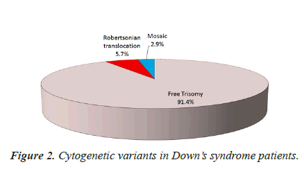
Out of 35 Down’s syndrome patients identified, a total of 32 (91.4%) had free trisomy. There were 2 (5.7%) cases with Robertsonian translocation and 1 (2.9%) having mosaic pattern (Table 2 and Figure 2).
| SN | Profile | No. | Percentage |
|---|---|---|---|
| 1. | Free Trisomy | 32 | 91.4% |
| 2. | Robertsonian translocation | 2 | 5.7% |
| 3. | Mosaic | 1 | 2.9% |
Table 2: Cytogenetic variants in Down’s syndrome Patients (n=35).
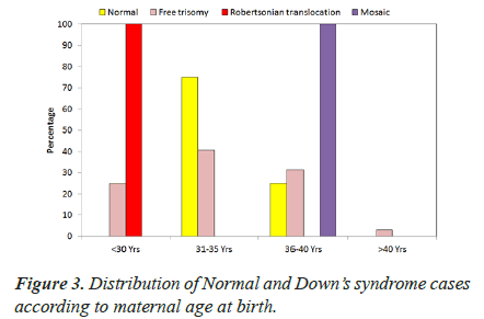
Maximum (n=13; 40.6%) cases of free trisomy had maternal age at birth in 31-35 years range followed by 36-40 years (n=10; 31.3%), < 30 years (n=8; 25%) and 1 (3.1%) in >40 years age range (Table 3 and Figure 3).
| Age Group (Yrs) | Normal (n=4) |
Free Trisomy (n=32) |
Robert-sonian Trans-location (n=2) | Mosaic (n=1) | Total (n=39) | |||||
|---|---|---|---|---|---|---|---|---|---|---|
| No. | % | No. | % | No. | % | No. | % | No. | % | |
| ≤ 30 Yrs. | 0 | 0.0 | 8 | 25.0 | 2 | 100.0 | 0 | 0.0 | 10 | 25.6 |
| 31-35 Yrs. | 3 | 75.0 | 13 | 40.6 | 0 | 0.0 | 0 | 0.0 | 16 | 41.0 |
| 36-40 Yrs. | 1 | 25.0 | 10 | 31.3 | 0 | 0.0 | 1 | 100.0 | 12 | 30.8 |
| >40 Yrs. | 0 | 0.0 | 1 | 3.1 | 0 | 0.0 | 0 | 0.0 | 1 | 2.6 |
χ2=10.395 (df=9); p=0.319
Table 3: Distribution of Down’s syndrome according to Maternal age at birth (n=35)
In Robertsonian translocation, both the cases were born to mothers in < 30 years age range.
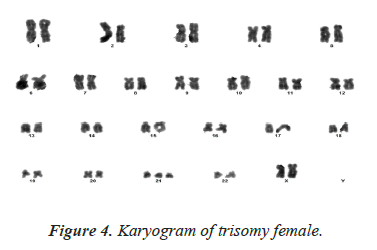
The lone case with mosaic pattern had maternal age at birth in 36-40 years range (Table 4) (Figure 4).
| Authors | Place | Free trisomy | Mosaicism | Translocation |
|---|---|---|---|---|
| Chandra et al. (2010) | Madras | 25.08 | 23.1 | 22.83 |
| El Gilany et al. (2011) | Egypt | 37.1 | 32 | 26.6 |
| Alois et al. (2013) | Mexico | 30 | 29 | 20 |
| Sotonica et al. (2016) | Sarajevo | 33 | 35.5 | 28 |
| Present study (2019) | Lucknow (India) | 33 | 38 | 25 |
Table 4: Comparative Analysis of Mean Maternal Age in DS Patients.
Discussion
In general, the overall incidence of Down’s syndrome is 1/800 live births, which seems to increase remarkably to 1/400 in mothers above 35 years of age, and to 1/12 by the age of 50 [9].
In the retrospective study conducted by Chandra et al. in Madras, the mean maternal age was 25.08 ± 4.77 for free trisomy cases, which was much higher than that for Robertsonian translocation cases (22.83 ± 3.89) and for mosaic individuals (23.1 ± 5.60) [10]. In another study by El Gilany et al. on Egyptian population, mothers of free trisomy cases were found to be older than mothers of translocation and mosaic cases (37.1 years vs. 26.6 and 32.0 years, respectively). Although this was in agreement with previous studies conducted on Egyptian population, [11] but vary from the data obtained in our study, to some extent, where mosaicism was better associated with advanced maternal age. Alois et al. observed that median of maternal age among free trisomy cases (30 years) and mosaic cases (29 years) was greater as compared to that in translocation cases (20 years) in Mexican population [12]. In our study, out of 32 cases of free trisomy, maximum (40.6%) had maternal age at birth in 31-35 years range followed by 36-40 years (31.3%), < 30 years (25%) and 3.1% in >40 years age range. The only case with mosaic pattern in our study had maternal age at birth in 36-40 years range. Therefore, we observed that advanced maternal age was more closely associated with mosaic variant followed by free trisomy. This finding was consistent with the study conducted by Sotonica et al. [7] who in their cross-sectional study, observed that the oldest mothers belonged to mosaic cases (35 years) followed by free trisomy cases (33 years) while youngest mothers belonged to translocation group of children (28 years) [7].
Both the cases having Robertsonian translocation in our study were born to mothers in < 30 years age range, which led to our conclusion that translocation variant was least associated with advanced maternal age. Chandra et al., El Gilany et al., Alois et al. and Sotonica et al., in their respective studies also observed translocation to be least associated with advanced maternal age group, compared to other variants, with mean maternal age of translocation group as follows: Chandra et al. -22.83 ± 3.89; El Gilany et al.-26.6 years; Alois et al.-20 years; Sotonica et al.-28 years. Our findings were thus in accordance with all the above studies.
On the other hand, Kolgeci et al. [13] in their cytogenetic study on DS cases in Prishtina, did not observe any effect of mother’s advanced age on prevalence of translocation variant of DS children [13].
Although most of the previous studies observed advanced maternal age as the principal risk factor for DS, but study conducted by Roy et al. [14] in Kolkata showed many DS babies born to the younger mothers (21-25 years of age) while only a few cases were associated with maternal age of >40 years [14]. The reason behind young mothers giving birth to more number of DS children is not clearly understood, but one of the possibilities could be the ovaries being biologically older than their real age in younger mothers [5]. Another contributory factor could be females getting married at an earlier age [15]. Some harmful practices like alcohol intake, tobacco smoking and drugs also seem to affect young females of reproductive age group very easily. Lesser hours of sleep, improper diet and unwanted pregnancies are some of the factors which are exclusively seen in younger mothers [7].
Conclusion
The most common cytogenetic variant of DS among the North Indian population is free trisomy. Advanced maternal age is one of the many risk factors associated with Down’s syndrome. In this study, advanced maternal age was seen to be more closely associated with mosaic variant followed by free trisomy. Translocation variant was least associated with advanced maternal age or we can say, it was associated with comparatively younger maternal age group. Limiting the number of pregnancies for mothers with advanced age can decrease the incidence of DS. Antenatal screening for DS, use of advanced medical tests for suspected cases and genetic counselling can help in lowering the burden of DS in society.
Acknowledgement
We wish to thank faculty and residents of Paediatrics Department for the permission to collect blood samples from the suspected patients. We would also like to thank Dr. Brijesh kumar, Dr. Priyanka Pandey and other seniors for their valuable suggestions and help.
References
- Kazemi M, Salehi M, Kheirollahi M. Down Syndrome: Current Status, Challenges and Future Perspectives. Int J Mol Cell Med Summer 2016; 5:126.
- Pande S, Salaskar V, Pais A, Pradhan G, Patil S, Parab C, Jadhav Y, MatkarS. Frequency of Down syndrome: An experience of a tertiary care diagnostic laboratory in India. Int J Adv Med 2017; 4: 1672-1675.
- Luz María Garduño- Zarazúa, Alois LG, Susana Kofman-Epstein, Alicia B. Peredo C. Prevalence of mosaicism for trisomy 21 and cytogenetic variant analysis in patients with clinical diagnosis of Down syndrome. Bol Med Hosp Infant Mex 2013; 70: 29.
- Coppede F. Risk factors for Down syndrome. Archive Fur Toxikologie 2016; 90: 1-10.
- Belmokhtar F, Belmokhtar R, Kerfouf A. Cytogenetic study of Down syndrome in Algeria: Report and review. J Med Sci 2016; 36: 46-52.
- Down’s Syndrome. National Down ’s syndrome Society.
- Sotonica M , Djurovic M, Hasic S, Kiseljakovic E, Jadric R, Ibrulj S. Association of parental age and type of Down’s syndrome on the territory of Bosnia and Herzegovina. Med Arch 2016; 70: 88-91.
- Plaiasu V. Down Syndrome-Genetics and Cardiogenetics. Maedica (Buchar) 2017; 12: 208–213.
- Murthy SK, Malhotra AK, Mani S, Shara MEA, Rowaished EM, Naveed S, AlKhayat AI, Al Ali MT. Incidence of Down Syndrome in Dubai, UAE. Med Princ Pract 2007; 16: 25–28.
- Chandra N, Cyril C, Lakshminarayana P, Nallasivam P, Ramesh A, Gopinath PM, Marimuthu KM. Cytogenetic Evaluation of Down Syndrome: A Review of 1020 Referral Cases. Int J Hum Genet 2010; 10: 87-93.
- Abdel-Hady El- Gilany, Yahia S, Shoker M, Faeza El- Dahtory. Cytogenetic and comorbidity profile of Down syndrome in Mansoura University Children's Hospital, Egypt. Indian J Hum Genet 2011; 17: 157–163.
- Luz María Garduño- Zarazúa, Alois LG, Susana Kofman-Epstein, Alicia B. Peredo C. Prevalence of mosaicism for trisomy 21 and cytogenetic variant analysis in patients with clinical diagnosis of Down syndrome. Bol Med Hosp Infant Mex 2013; 70: 29.
- Kolgeci S, Kolgeci J, Azemi M, Ruke Shala-Beqiraj, Gashi Z, Sopjani M. Cytogenetic Study in Children with Down Syndrome Among Kosova Albanian Population Between 2000 and 2010. Mat Soc Med 2013; 25: 131-135.
- Roy S, Tapadar A, Kundu R, Ghosh S, Halder A. Cytogenetic variations in a series of cases of Down’s syndrome. Journal of the Anatomical Society of India 2015; 64: 73-78.
- Malini SS, Ramachandra NB. Influence of advanced age of maternal grandmothers on Down Syndrome. BMC Med Gen 2006; 7: 4.


