ISSN: 0970-938X (Print) | 0976-1683 (Electronic)
Biomedical Research
An International Journal of Medical Sciences
Case Report - Biomedical Research (2018) Volume 29, Issue 10
Bilateral congenital dacryocystocele-A case report
DOI: 10.4066/biomedicalresearch.29-18-388
Visit for more related articles at Biomedical ResearchCongenital dacryocystocele is a rare congenital anomaly of the lacrimal drainage system (0.02% of newborns), which refers to the cystic dilatation of the lacrimal sac and nasolacrimal duct. In most cases the pathology is unilateral with female predominance. Bilateral cases constitute 10% of all cases and in most babies with bilateral disease there are coexistent nasal cysts. Clinically, dacryocystocele presents as a grey-bluish cystic swelling of the lacrimal sac in the medial canthal area that typically present at birth and often resolve spontaneously with conservative management in the first months of life. Bilateral cases of congenital dacryocele are rarely reported, especially isolated dacryocystoles without coexistent nasal cysts. We present a rare case of bilateral congenital dacryocystocele in a 10-day-old newborn boy, without concomitant intranasal cysts. The infant was treated conservative with spontaneous resolution of the disease 6 weeks after treatment initialization. No dacryocystitis or preseptal cellulitis was observed, nevertheless rupturing of the dacryocystocele to the common canaliculus.
Keywords
Bilateral, Congenital, Dacryocystocele, Conservative treatment, Spontaneous resolution
Introduction
Congenital dacryocystocele is a rare congenital anomaly of the lacrimal drainage system which refers to the cystic dilatation of lacrimal sac and nasolacrimal duct often extending into the inferior meatus and further into the nasal passage [1]. In most cases it is unilateral condition and predominantly occurs in girls [2]. Clinically, dacryocystocele presents as a grey-bluish cystic swelling of the lacrimal sac in the medial canthal area that typically present at birth [3,4] and often resolve spontaneously with conservative management in the first months of life [5]. Computed tomography and magnetic resonance imaging are performed in differential diagnostic difficult cases when meningoencephalocele is suspected [4]. In all cases with bilateral dacryocystocele, a careful nasal examination must be done for ruling out coexistent nasal cysts that can cause sudden respiratory distress syndrome in neonates [4]. Bilateral cases of congenital dacryocele are rarely reported, especially isolated dacryocystoceles without coexistent nasal cysts. We report a rare case of a 10-day-old newborn boy with bilateral congenital dacryocystocele without coexistent nasal cysts.
Case Report
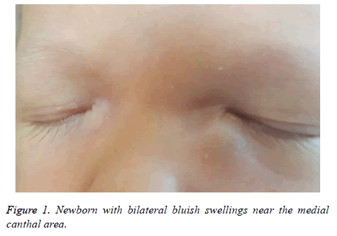
A 10-day-old newborn boy was presented to our outpatient care with bilateral bluish swellings near the medial canthal area (Figure 1). The infant was born mature, without any history of other congenital pathology. Parents noticed the bluish swellings on the third day after delivery, but no treatment had been started. There was no history of tearing or inflammation symptoms. The swelling was more obvious on the left side and two masses were tender without fluid reflux from the lacrimal puncti.
The baby was diagnosed with bilateral congenital dacryocystocele and otorhinolaryngologist consultation with endoscopy examination was made that ruled out intranasal cystic extension. A conservative treatment with gentle sac massage and warm compresses was prescribed. A close follow up (every 1 week) of the baby was initiated. Rupture of the dacryocystocele to the common canaliculus with mucoid fluid reflux in both eyes was observed on 10th day after treatment initialization. Topical antibiotic was additionally prescribed and sac massage and warm compresses continued. No signs of secondary dacryocystitis or preseptal cellulitis were observed. Recurences of the swellings of both eyes was observed for a period of six weeks, after that full resolution of the sac masses was recorded just with conservative treatment. The patient has been followed up for two months without any signs of nasolacrimal duct obstruction.
Discussion and Conclusion
Dacryocystocele is a rare congenital condition that can be observed in 0,005% of newborns [6]. A double obstruction of the lacrimal system distally and proximally to the lacrimal sac during fetal life is responsible for cystic dilation of lacrimal pathway [1]. The obstruction is anatomical at the level of Hasner valve and relative (functional) at the level of the common canaliculus (Rosenmuller valve) [3]. Because of the relative obstruction of the Rosenmuller valve amniotic fluid, mucus or tears can enter the lacrimal sac but can’t drain upwards through the common canaliculus because of the Rosenmuller valve collapse or downwards because of the anatomical barrier of the Hasner valve, forming sac swelling [3]. In most cases dacryocystocele is a unilateral condition (~90%) and have strong female preponderance (~70%) [2]. Clinically, congenital dacryocystocele presents as an enlarged grey-blue cystic swelling in the medial canthal area, epiphora, and high tear meniscus height [2-4,7] at or shortly after birth [4]. The differential diagnosis is very broad and includes encephalocele, meningoencephalocele, capillary haemangioma, dermoid cyst, lymphangioma, nasal glioma and etc [2,4]. Ultrasonography is very important non-invasive method that can be used pre and postnatally for the diagnosis and differential diagnosis [1,4,8], as dacryocystocele is seen as a hypoechogenic mass located inferiomedially with ostium connected with the dilated nasolacrimal duct that may be seen in the coronal or parasagittal plane including the nose and medial angle of the orbits [1,4]. The ultrasonographic diameter of the cyst varies with mean size of 11.5 mm in 5 neonates with congenital dacryocystocele reported by Cavazza et al. [4]. Whenever cranial pathology is suspected (meningocele, meningoencephalocele) computed tomography or magnetic resonance imaging must be performed [1,4]. In all cases with dacryocele and especially in cases with bilateral disease intranasal cyst must be ruled out with nasal bilateral endoscopy [4,9]. The nasal cysts may be a direct extension of the nasolacrimal duct, located beneath the inferior turbinate [9] and can be responsible for 6% of cases with congenital nasal obstruction [10]. Neonates are obligatory nasal breathers therefore nasal cyst can be reason for acute respiratory distress syndrome [4], especially during feeding and sleeping.
Management of dacryocystoceles remains controversial [4]. Some authors advocate for conservative treatment with sac massage, warm compresses and topical antibiotic drops with two weeks resolution in 76% of patients [5] and surgical treatment (probing under general anaesthesia) in the first months of life after non-resolution of the cyst [4] or urgent surgery in cases with dacryocystitis, cellulitis, large cyst that causes astigmatism or narrowing of the lid fissure and especially in patients with intranasal cyst and respiratory dyspnea [3,4]. Other physians prefer early surgical treatment because of the high risk of dacryocistitis development [7]. In cases with nasal obstruction, probing as an isolated surgical manner is not sufficient and concomitant extensive marsupialization of the dacryocystocele must be performed [2,3].
References
- Bingöl B, Basgül A, Güdücü N, Isci H, Dünder I. Prenatal early diagnosis of dacryocystocele, a case report and review of literature. J Turk Ger Gynecol Assoc 2011; 12: 259-262.
- Shashy RG, Durairaj VD, Holmes JM. Congenital dacryocystocele associated with intranasal cysts: diagnosis and management. Laryngoscope 2003; 113: 37-40.
- Kim H, Park J, Jang J, Chun J. Urgent bilateral endoscopic marsupialization for respiratory distress due to bilateral dacryocystitis in a newborn. J Craniofac Surg 2014; 25: 292-293.
- Cavazza S, Laffi GL, Lodi L, Tassinari G, Dall’Olio D. Congenital dacryocystocele: diagnosis and treatment. Acta Otorhinolaryngol Ital 2008; 28: 298-301.
- Schnall BM, Christian CJ. Conservative treatment of congenital dacryocele. J Pediatr Ophthalmol Strabismus 1996; 33: 219-222.
- Davies R, Watkins WJ, Kotecha S, Watts P. The presentation, clinical features, complications, and treatment of congenital dacryocystocele. Eye 2018; 32: 522-526.
- Becker BB. The treatment of congenital dacryocystocele. Am J Ophthalmol 2006; 142: 835-838.
- Davis WK, Mahony BS, Carroll BA, Bowie JD. Antenatal sonographic detection of benign dacrocystoceles (lacrimal duct cysts) J Ultrasound Med 1987; 6: 461-465.
- Grin TR, Mertz JS, Stass-Isern M. Congenital nasolacrimal duct cysts in dacryocystocele. Ophthalmol 1991; 98: 1238-1242.
- Patel VA, Carr MM. Congenital nasal obstruction in infants: A retrospective study and literature review. Int J Pediatr Otorhinolaryngol 2017; 99: 78-84.
