ISSN: 0970-938X (Print) | 0976-1683 (Electronic)
Biomedical Research
An International Journal of Medical Sciences
- Biomedical Research (2013) Volume 24, Issue 2
Bilateral variations in the growth and development of human foetal clavicle.
Being a long bone and having an intramembranous ossification with earliest appearance of primary ossification (two) centres and having no medullary cavity it reflects that nature too supports its importance in gaining early strength so that it can support the developing upper limbs of the foetus earliest to provide them easy movements, our study is to determine the bilateral growth variations during the intra uterine life. 60 clavicles were obtained from 30 human foetuses ranging from 14 weeks to 33 weeks of IUL from Department of Anatomy, Jawaharlal Nehru, Aligarh Muslim University, Aligarh. For the purpose of study, foetuses were divided into five groups on the basis of gestational age. Parameters selected to determine the bilateral variations were weight; length; circumference, AP and Vertical diameter at midshaft; AP and Vertical diameter at medial end; AP and Vertical diameter at lateral end of clavicles. All parameters were measured using vernier calliper and weight was taken by the single pan fractional weighing mechine. Students t test was used to determine the coefficient of significance, SPSS Software was used to do the calculations. Later linear graphs were prepared considering both right and left clavicles for the same parameter. It was found that bilateral variations were not significant in most of the parameters considered in our study, except thickness of its shaft at its midpoint which is more pronounced on the right side in early foetal life and on the left side in late foetal life. Thus we conclude that there is no significant bilateral variations in growth and development of clavicle during the intrauterine life. The variations that may be present in the adult clavicles are due to the factors that play their role after birth.
Keywords
Morphometry, bilateral variations, clavicle, human foetus, clavicular length
Introduction
The developmental anatomy is gaining increasing significance, as it constitutes the basic framework of different clinical specialties possessing a foetal, neonatal or paediatric orientation. In fact the morphology of an organ often sufficiently tells the practicing physicians more than many functions. Hence, there is a continuing need for morphological data.
A lot of work has been done till date to assess the age of the foetuses (in utero), Schwarzler [1] prepared Sexspecific antenatal reference growth charts for uncomplicated singleton pregnancies at 15-40 weeks of gestation. Persson [2] assessed the reliability of ultrasound fetometry in estimating gestational age in the second trimester. Natalie [3] compared the age and sex estimation of clavicle by traditional and novel methods. Shobha [4] determined sex of adult human clavicles by morphometric parameters. However till date no study is avalable to assess the age of a dead foetus or of foetal remains which may be useful for medico legal purpose.
Many morphometric studies have been done on long bones of foetuses, Lowrance [5] and Khan and Faruqi [6] on Asian subjects; Nasrat and Bondagji [7] worked on ultrasound biometry of Arabian foetuses. Sherer [8] worked on the foetal clavicle length throughout gestation by means of ultrasonography, Yarkoni [9] found that how the clavicular measurements can be a new biometric parameter for foetal evaluation. Diaphyseal lengths of dried material of foetal skeletons from the third to the tenth Lunar month of pregnancy have already been investigated in a forensic series by Fazekas and Kosa [10] but it lacked information about human foetal clavicle.
All these studies were based on radiographic or sonographic evaluation. Frutos [11] worked for determination of sex from clavicle and scapula in a Guatemalan contemporary rural indigenous population and by means of actual manual measurements but his work was on adult clavicles, Odgen [12] worked on 31 pairs of adult human clavicles from human cadavers (fullterm stillborn to fourteen years) by means of radiology to know their post natal development. No work has been done in the field of morphometry on human foetal clavicle in actual means, because in every aspect manual measurements will give the most precise data than by radiography or by sonography so our results will be the most accurate ones in the field of morphometry of human foetal clavicle till date.
Being a long bone and having an intramembranous ossification with earliest appearance of primary ossification (two) centres and having no medullary cavity it reflects that nature too supports its importance in gaining early strength so that it can support the developing upper limbs of the foetus earliest to provide them stable movements. Moore [13]. The clavicle varies more in shape than most other long bones, it’s thicker and more curved in manual workers and the sites of muscular attachments are more marked.
Taking into account the aforementioned arguments, our study is to ascertain wether there are any bilateral variations found in the growth and development of clavicle during the intrauterine life.
Materials and Method
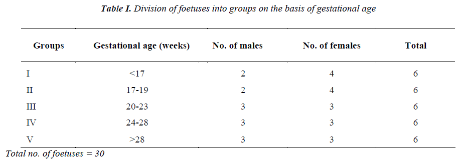
30 (13 male and 17 female) Human Foetuses were obtained from the Museum, Department of Anatomy, Jawaharlal Nehru Medical College, A.M.U. Aligarh after being awarded Ethical Clearance Certificate from the Institutional Ethics Committee (IEC). Foetuses of all age groups without congenital craniovertebral anomalies (e.g. anencephaly, spina bifida, cleidocraniodysostosis) were selected for the study. The parameter used for determination of gestational age was foetal foot length. Which is already been documented by Streeter [14]. For the purpose of study, foetuses were divided into five groups on the basis of gestational age.
Determination of sex was done taking into consideration the external genitalia.
Measurements taken
1. Weight (mm)
2. Length (mm)
3. Circumference at the midshaft (mm)
4. Anteroposterior diameter at the midshaft (mm)
5. Vertical diameter at the midshaft (mm)
6. Anteroposterior diameter at the medial end (mm)
7. Vertical diameter at the medial end of both left and right clavicles (mm)
8. Anteroposterior diameter at the lateral end (mm)
9. Vertical diameter at the lateral end of both left and right clavicles (mm)
All parameters were measured using vernier calliper and weight was taken by the single pan fractional weighing mechine. Student’s t test was used to determine whether statistically significant differences occur between the different measurements taken from individual bones, both right and left clavicles from 30 (13 male and 17 female) human foetuses. SPSS software was used to solve the calculations.
Results
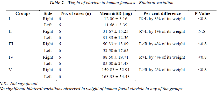
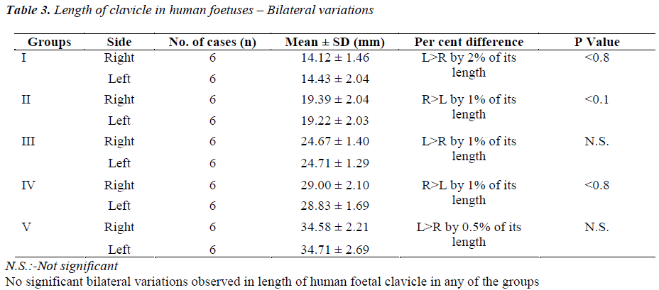
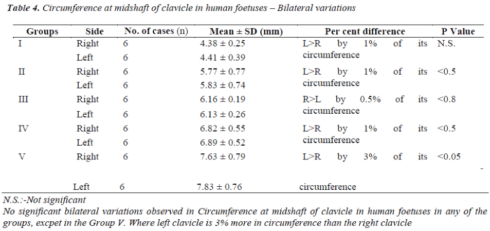
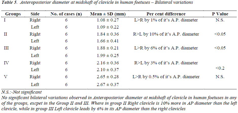
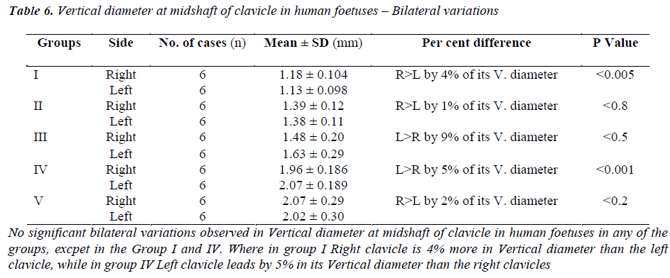
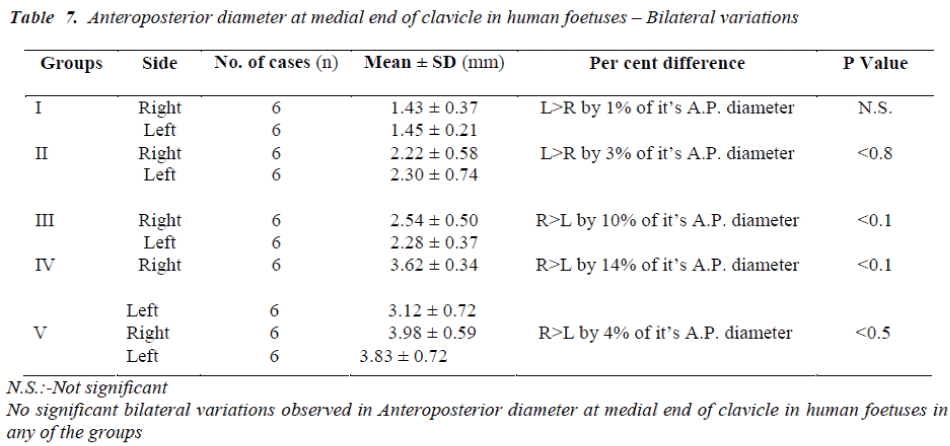
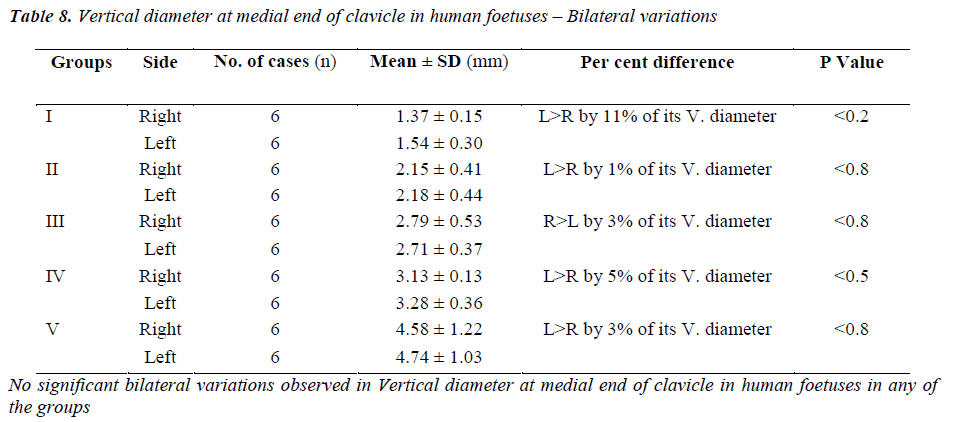
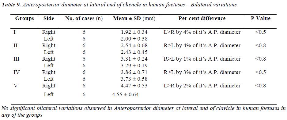
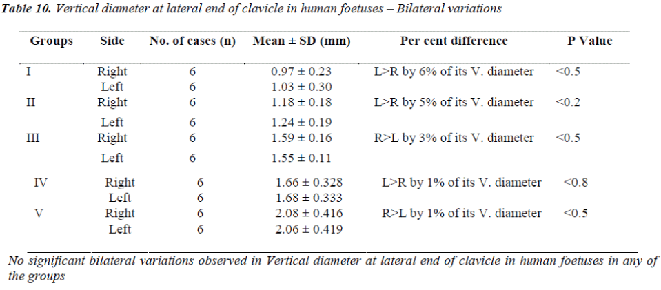
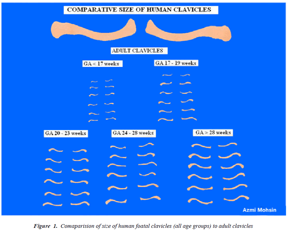
All the parameters were measured on both the right and left clavicles in each age group and mean of the group with its standard deviation was taken into consideration, P Value and Percentage difference were calculated (Tables 2-10, (Fig. 1).
Discussion
Regarding Bilateral variations, Bagnall [15], presented a complicated picture of growth of the two sides of the foetal body, from his study he could not conclude any inference regarding right or left dominance of skeletal growth during foetal life. Similarly in our study bilateral variations are not noticed in most of the parameters considered in our study. Midshaft readings including diameters and circumference are the only parameters which show some significant variations but in few groups of foetuses only. The anteroposterior diameter at the midshaft of clavicle shows significant bilateral variation (p <0.05) in group II & III only (Table. V). in the former the value in right clavicle is more while in latter the reading in the left clavicle is greater. Vertical diameter at midshaft of clavicle shows significant variation in group I (p <0.005) and group IV (p <0.001) (Table. VI), here also we find that smaller clavicle shows right dominance while the larger clavicle shows left dominance. As far as circumference at midshaft is concerned there is significant bilateral variation only in group V specimens (p <0.05) (Table. IV). interestingly here also the value was more in the left clavicle. So we conclude that parameters of the midshaft of clavicle show right dominance in early foetal life and left dominance in late foetal life, which is of no significance as it is noticed only around the mid shaft while all other parameters show no significant bilateral variations. However in adulthood the right clavicle is usually stronger and shorter than the left clavicle. Moore [13]. We further conclude that, right dominance in the growth and development of clavicle in most of the right handed population seem to be an acquired phenomenon. Benjamin and Michelle [16].
Conclusion
Growth and development of clavicle during intra uterine life is nearly a symmetrical phenomenon, Whatever Bilateral variations observed in adult clavicle are due to factors which play their role after birth.
References
- SchwarzlerP, Bland JM, Holden D, Campbell S. Ville Y. “Sex-specific antenatal reference growth charts for uncomplicated singleton pregnancies at 15-40 weeks of gestation.” Fetal Medicine Unit and Department of Public Health Sciences, St. George’s Hospital Medical School, London, UK. ISUOG. John Wiley & Sons, Ltd. 2004.
- Persson PH and Weldner BM. “Reliability of ultrasound fetometry in estimating gestational age in the second trimester.” Acta Obstet Gynecol Scand 1986; 65 (5): 481-483.
- Shirley NR. Age and sex estimation from the human clavicle: An investigation of traditional and novel methods. This report has not been published by the U.S. Department of Justice. To provide better customer service, NCJRS has made this Federally-funded grant final report available electronically in addition to traditional paper. Document No.: 227930, Date Received: (August 2009), Award Number: 2007-DNBX- 0004
- Shobha “Determination of sex of adult human clavicle by morphometric parameters” dissertation submitted to the Rajiv Gandhi University of Health Sciences, Banglore, Karnataka. India (2010).
- http://119.82.96.198:8080/jspui/bitstream/123456789/4836/1/Shobha.pdf
- Lowrence EW, Homer B, Latimer “Measurements of human bones from Asia,” Transactions of the Kansas Academy of Science 1964; 67 (1); 25-30.
- Khan Z, Faruqi NA. Determination of Gestational Age of Human Foetuses from Diaphyseal lengths of long bones – A radiological study.” Journal of Anatomical Society of India 2006; 55 (1): 67-71.
- Nasrat H, Bondagji NS. Ultrasound biometry of Arabian fetuses. Int J Gynaecol Obstet 2005; 88(2): 173-178. Epub 2004 Dec 30.
- Sherer DM, Sokolovski M, Dalloul M, Khoury-Collado F, Osho JA, Lamarque MD. Abulafia O. Fetal clavicle length throughout gestation: a normogram.” Ultrasound Obstet Gyneco. 2006; 27(3): 306-310.
- Yarkoni S., Schmidt W., Jeanty P., Reece E. A. and Hobbins J. C. “Clavicular measurement: a new biometric parameter for fetal evaluation.” Journal of Ultrasound in Medicine 1985; 4 (9): 467-470.
- Fazekas I. and Kosa K. “Forensic fetal osteology”, Budapest Hungary Akademiai Kiado Publishers, 1978, 232-277.
- Frutos LR. Determination of sex from the clavicle and scapula in a Guatemalan contemporary rural indigenous population”, The American Journal of Forensic and pathology (Am. j Forensic Med. Pathol ISSN 0195- 7910 2002; 23 (3) 284-288.
- Ogden JA, Conlogue GJ, Bronson ML. Radiology of postnatal skeletal development. III. The Clavicle. Skeletal Radiol 1979; 4 (4): 96-203.
- Moore KL and Dalley AF. Variations of the clavicle.” Clinically Oriented Anatomy 2006;, 5th ed. P729.
- Streeter GL. Weight, sitting height, head size, foot length & menstrual age for human embryo. Contribution to Embryology 1920; 11: 143.
- Bagnall KM, Harris PF, Jones PRM. A radiographic study of the longitudinal growth of primary ossification centres in limb long bones of the human fetus. The Anat Rec 1982; 203: 293-299.
- Benjamin M. Auerbach, Michelle H. Raxter “Patterns of clavicular bilateral asymmetry in relation to the humerus: variation among humans” Journal of Human Evolution 2008; 54: 663 e 674
