ISSN: 0970-938X (Print) | 0976-1683 (Electronic)
Biomedical Research
An International Journal of Medical Sciences
Research Article - Biomedical Research (2017) Volume 28, Issue 20
Biomechanical analysis of discus athletes' acute injury of shoulder joint at the beginning phase of rotation
Zhikun Hou*
Department of Physical Education, Shaanxi University of Chinese Medicine, Xi’an, 712046, PR China
- *Corresponding Author:
- Zhikun Hou
Department of Physical Education
Shaanxi University of Chinese Medicine, PR China
Accepted on October 28, 2017
To further clarify possible sports injury of shoulder joint of excellent discus athletes at beginning phase of rotation and biomechanic characteristics demonstrated, the paper adopts close range dynamic stereo photography measurement method for fixed-point shooting of live competition, training of discus athletes. Comprehensive analysis and research of motion image and information is done with professional video, finally biomechanical indexes of athletes are obtained, and in-depth inquiry is done for possible sports injury of shoulder joint at beginning phase of rotation and biomechanical characteristics demonstrated. Results show that the highest constituent ratio of shoulder joint injury of excellent discus athletes comes from injury of tendon of long head of biceps brachii, followed by rotator cuff injury, acromioclavicular joint sprain and dislocation, dislocation and subluxation (glenohumeral joint), deltoid muscle strain. Biomechanical results show that biomechanical indexes of shoulder joint at beginning phase of rotation are as follows: right from the moment of lifting of right foot, shoulder and hip angle is 45 degrees, pulling angle is 126 degrees, shoulder axis angle is 2 degrees, velocity of left shoulder is 2.64 m/s, velocity of right shoulder is 0.95m/s; right from the moment of lifting of left foot, shoulder and hip angle is 27 degrees, pulling angle is 150 degrees, shoulder axis angle is -12 degrees, speed of left shoulder is 1.96 m/s, speed of right shoulder is 1.97 m/s. In conclusion, at beginning phase of rotation, increased pulling angle caused the highest ratio of injury of tendon of long head of biceps brachii. Therefore, in the daily training and competition, adjusting of corresponding angle should be a focus.
Keywords
Discus movement, Shoulder joint, Acute injury, Biomechanical analysis.
Introduction
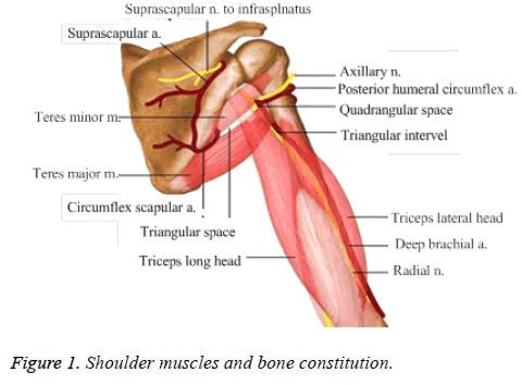
Biomechanics is a biophysics branch of quantitative research of biology mechanics problems with principles and methods of mechanics. Its research scope ranges from biological whole to the system, organs (including blood, body fluids, organs, bones, etc.), from bird flying, fish swimming, flagellum and ciliary movement to transportation of plant body fluid, etc. The basis of biomechanics is the law of energy conservation, law of momentum, three laws of mass conservation and constitutive equation describing physical properties [1-3]. Biomechanics mainly applies physical laws and engineering concepts to describe the relationship between dynamic and force. Weiss et al. showed that biomechanics was associated with patellofemoral pain and anterior cruciate ligament (ACL) injuries in sports [4]. Discus sport is a main type of throwing with very common sports injury problems, mainly muscle ligament sprain or fracture due to insufficient preparation activities at training and competition or unskilled techniques [5]. A study showed an analysis of biomechanics on the sports injury of the knee joint at stage of high rising transition in male discus athletes [6]. Another research on the technology training of discus throw based on the sports biomechanics have beens also reported [7]. In addition, as technical characteristics can also cause fatigue damage, throwing events often need extraordinary range of abnormal joint activities. Kinematics can provide quantitative description of joint activities range, provide scientific basis for avoiding damage and changing sports rule, and provide basis for improvement of protective equipment. The shoulder is the body’s most mobile joint, and injury of discus athletes largely occurs in the shoulder joint [8,9] (Figure 1). However, the biomechanical analysis of discus athletes’ acute injury of shoulder joint was limited. Therefore, in this study, we aimed to further clarify possible sports injury of shoulder joint of excellent discus athletes at beginning phase of rotation. In order to provide more reasonable training methods to excellent discus athletes and improve performance, this paper conducts in-depth analysis of shoulder joint injury problems of discus athletes from point of view of biomechanics with specific circumstances.
Method
General information
Subjects: 400 excellent discus athletes during March 2012 - March 2015, with 230 male athletes, 174 female athletes were enrolled in this study. Follow-up survey with 404 questionnaires was issued every year and 1212 questionnaires were issued in three years.
Questionnaire survey
The main contents of the questionnaire include cause of shoulder joint, nature of injury and injury site, to understand constituent ratio of shoulder joint injury of discus athletes.
Measurement method
In June 2014, close range dynamic stereo photography (Company in China) measurement method is taken for fixedpoint shooting of athletes’ live match and training, with shooting frequency regulated at 50 Hz [10]. Motion video rapid feedback analysis system is adopted to fulfill image sampling and data calculation.
Biomechanical indexes
Indexes of shoulder and hip angle, pulling angle, shoulder axis angle, velocity of left shoulder, velocity of right shoulder from the moment of left foot touchdown to disposing of discus.
Observation index
Constituent ratio of discus athletes’ shoulder joint injury at stage of final exertion, indexes of shoulder and hip angle, pulling angle, shoulder axis angle, velocity of left shoulder, velocity of right shoulder from the moment of left foot touchdown to disposing of discus. The inclusion criteria: all participants were discus athletes.
Statistical methods
SPSS19.0 statistics software is used to complete data entry and output, with average number ± mean value to denote measurement data and t value for test.
Ethical consideration
The study was carried out in compliance with the Declaration of Helsinki of the World Medical Association, and according to a protocol approved by Shaanxi University of Chinese Medicine, the approval number is 2012009. The objectives of the study were explained to the study participants and verbal consent was obtained before interviewing each participant.
Results
The 1212 questionnaires are issued to participants based on quantitative analysis, with all recovered, with efficiency at 100%. Data result of 1212 questionnaires are simultaneously analyzed by investigation team. Subjects were on average of (21.43 ± 2.95) years old. Based on shoulder joint injury nature and injury site of 400 excellent discus athletes, the highest constituent ratio of shoulder joint injury comes from tendon of long head of biceps brachii, 34.65% (420/1212); followed by rotator cuff injury, 23.76% (288/1212); acromioclavicular joint sprain and dislocation, dislocation and subluxation (glenohumeral joint), deltoid muscle strain and other injuries constitute 11.88% (144/1212), 1.98% (24/1212), 7.92% (96/1212), 19.81% (240/1212).
Refer to the following table for relation between shoulder joint angle and speed change from the moment of lifting of right foot to the moment of lifting of left foot of 400 excellent discus athletes. The shoulder joint angle and speed change from the moment of lifting of right foot to the moment of lifting of left foot is shown as Table 1.
| Timing | Shoulder hip (degrees) | Pulling angle (degrees) | Shoulder axis angle (degrees) | Velocity of left shoulder (m/s) | Velocity of right shoulder (m/s) |
|---|---|---|---|---|---|
| The moment of lifting of right foot | 44 ± 1 | 127 ± 6 | 2 ± 1 | 2.64 ± 1.21 | 0.99 ± 0.41 |
| The moment of lifting of left foot | 26 ± 5 | 150 ± 3 | -11 ± 3 | 1.97 ± 0.33 | 1.92 ± 0.73 |
Table 1. Shoulder joint angle and speed change from the moment of lifting of right foot to the moment of lifting of left foot.
Musculus biceps brachii is one of the most important upper limb muscles. Tendon of biceps brachii connects musculus biceps brachii and bone. Its role includes: elbow flexion, supination of forearm, lower head of humerus, shoulder flexion. Musculus biceps brachii is divided into two tendon of caput longum and brachycephaly at shoulder joint. Caput longum goes over the superior humeral head and is attached to glenoid, the part most likely to cause symptoms. Musculus biceps brachii and musculus triceps brachii together constitute main part of upper arm, muscle group located in the front of the upper arm. In terms of its function, mainly flexion of the arm exerts to complete all pulling action and assist in activities of muscle group at the back towards the outside world.
Rotator cuff is a group of tendon complex wrapped around humeral head. The front of humeral head is subscapularis tendon, upward side is supraspinatus tendon, the rear part is infraspinatus tendon and teres minor tendon. Movement of these tendons results in shoulder internal rotation, external rotation and lifting activities, but the more important is that these tendons stabilize humeral head in the glenoid, which plays an extremely important role in maintaining shoulder joint stability and activity of shoulder joint.
Shoulder girdle is composed of 5 joints, namely glenohumeral joint, acromioclavicular joint, sternoclavicular joint, joint between the chest wall and the shoulder and acromion humeral joint. Therefore, any joint injury will affect shoulder movement function in varying degrees. The shoulder joint is with maximum human body activity. Because of small size of glenoid, and as humeral head is big and round, joint capsule is relatively loose, glenohumeral joint is with high activity, which is different from hip. Plus rise and fall, rotation of scapula and its orbiting (adduction and abduction) along the chest wall, scope of activities is greater. Therefore, during movement, the shoulder can complete a wide range of relatively complex actions and thus is prone to hurt. Instant action from lifting of right foot to lifting of left foot is to begin rotation for the sake of body stability, increase moment of inertia, reserve energy and create favorable conditions for rotational speed at flight.
Discussion
Our results showed that at the moment of lifting of right foot, mean shoulder and hip angle of excellent discus athletes is 45 degrees, mean pulling angle is 125 degrees. At this time, athletes’ body postures gradually expand through hip twisting from maximum tightening state at preliminary swing, with body tightening weakened. At the moment of lifting of right foot, mean shoulder and hip angle is 26 degrees, mean pulling angle is 150 degrees. During the moment from lifting of right foot to lifting of left foot, shoulder and hip angle becomes smaller, pulling angle becomes larger, the body gradually expands with right shoulder turning with the left side, and pulling angle is increased by an average of 23 degrees at this stage.
Pulling angle increase of excellent discus athletes is relatively large at this stage. Athletes’ left shoulder extends in the throwing direction, with right arm passively rolling forward at the back. The right shoulder excessively extends, rotates and stretches, velocity of right shoulder increases while velocity of left shoulder declines. The front structure of right shoulder is in passive tension and tension. Acromioclavicular joint is formed of inside end of acromion, outer end of clavicle linked with joint capsule, acromioclavicular, ligament, deltoid, trapezius muscle tendon attachment and coracoclavicular ligament [11,12]. Acromioclavicular joint function is demonstrated in lifting and fall of scapula, and scapular adduction and abduction. With increase in amplitude of variation of pulling angle, throwing arm fails to turn in throwing direction with body, but exerts backward, which may be an important factor in occurrence of acromioclavicular joint sprain and dislocation. To try to avoid acromioclavicular joint sprain, shoulder girdle should be appropriately relaxed, throwing arm should turn in throwing direction with body, without active exertion backward, which is also a technical requirement to improve athletic performance. The results show that the highest constituent ratio of sports injury of national master athletes comes from injury of tendon of long head of biceps brachii, 35%. Caput longum of musculus biceps brachii begins from glenoid nodule joint, while tendon descends in joint, passes through intertubercular groove and connects to vaginae synoviales with joint capsule. It is the only tendon through joint in the body. Intertubercular sulcus is narrow, with transverse ligament protection to prevent tendon dislocation. The brachycephaly begins from coracoid, while two muscle belly gradually integrate into one, separate into two in the downside, respectively ending at tuber radii and medial forearm fascia. In addition to elbow flexion and rear suspension of forearm, musculus biceps can stabilize shoulder joint and extend humeral at elbow suspension [13]. By questionnaire survey and three-dimensional biomechanical analysis of sports, Li found that the sports injury and biomechanical characteristics of shoulder joint that may appear in the state outstanding women discus athletes at the beginning phase of rotation, which was consistent with this study [14]. In addition, another study also reported an analysis of biomechanical on the acute injury of shoulder joint of finally forcibly phase in male discus athletes [15]. Due to relatively large amplitude increase of pulling angle at this stage, the highest constituent ratio of injury comes from tendon of long head of biceps brachii, and increased amplitude of pulling angle may be an important factor that causes injury of tendon of long head of biceps brachii. Athletes are thus required to pay attention to relaxation of the right shoulder girdle after completion of action. Although the bigger pulling angle is, the better, the limit should not be overrun to avoid injury of tendon of long head of biceps brachii. Questionnaire survey results show that mean shoulder axis angle of national master athletes at the instant moment of lifting of left foot is -12°; at this moment, included angle of shoulder and hip axis and perpendicular plane in throwing direction is located at left front of throwing circle, while hip axis precedes shoulder axis and turns to right front of throwing circle.
References
- Yuanjing L. Biomechanical analysis of discus athletes’ acute injury of shoulder joint at the beginning phase of rotation. Chinese J Tissue Eng Res 2007; 11: 3341-3343.
- Wenmei D, Yuanjing L, Yanxia W. Biomechanics analysis of acute injury of shoulder joint of male discus athletes at the stage of final exertion. Chinese J Clin Rehab 2005; 9: 116-117.
- Jinguang Y. Research of sports representation training method of juvenile discus athletes. Contemporary Sports Technol 2013; 3: 44-46.
- Weiss K, Whatman C. Biomechanics Associated with Patellofemoral Pain and ACL Injuries in Sports. Sports Med 2015; 45: 1325-1337.
- Tianwei L. Kinematics analysis of discus technology of chinese elite female discus athlete Li Yanfeng. Xi’an Physical Education University, 2012.
- Yuan-Jing LI, Chen J, Wei-Long XU. Biomechanics analysis on the sports injury of the knee joint at stage of high rising transition of male discus athletes. J Guangzhou Sport University 2016l; 23: 256-257.
- Cui DX, Ai-Guo LU. Research on the technology training of discus throw based on the sports biomechanics. J Hebei Institute Physical Edu 2011; 16: 121-123.
- Dianchen J. Investigation and analysis of strength training of juvenile discus athletes in Tonghua city. Modern Commun 2014; 42: 130.
- Liang Z. Rhythm characteristics analysis of motion of center of gravity of chinese excellent female discus athletes at the stage of final exertion. J Tianjin Uni Sport 2009; 24: 265-268.
- Xiaoqiang N, Limin W. Comparative analysis of kinematics of male discus athletes at home and abroad. Value Eng 2014; 11: 325-326.
- Ni M, Liying X. Biomechanical analysis in design of individualized artificial knee joint based on LifeMOD. Chinese J Rehab Med 2011; 26: 538-542.
- Tao L, Jiajun Z, Tingting W. 3D Finite element simulation and biomechanical analysis of mandibular symphysis explosion injury. Chinese J Trauma 2015; 31: 1050-1055.
- Lin L, Huanbin Z, Yi S. Biomechanical analysis of back gliding shot put technique of female shot putters. J Shandong Institute Physical Edu Sports 2008; 24: 76-79.
- Li YJ. Biomechanical analysis of acute injury of the shoulder joint in discus athletes at the beginning phase of rotation. J Clin Rehab Tissue Eng Res 2007.
- Dong WM, Li YJ, Wu YX. Biomechanical analysis on the acute injury of shoulder joint of finally forcibly phase in male discus athletes. Chinese J Clin Rehab 2005; 9: 116-117.
