ISSN: 0970-938X (Print) | 0976-1683 (Electronic)
Biomedical Research
An International Journal of Medical Sciences
- Biomedical Research (2009) Volume 20, Issue 2
Bowel perforation secondary to mesenteric carcinoid tumor: A case report and review of the literature
Carcinoid tumor is rare compared to other tumors of the gastrointestinal tract. Mesenteric carcinoid tumor is almost always due to a metastasis from a carcinoid tumor of the small bowel beyond the ligament of Treitz. CT remains the dominant imaging modality for the diagnosis of mesenteric neoplasms. We present an unusual case of mesenteric carcinoid with small bowel perforation.
Keywords
Mesenteric carcinoid, bowel perforation
Introduction
Primary tumors arising from the mesentery are rare. Most primary lesions are mesenchymal in origin, and the majority is histologically benign. Gastrointestinal carcinoid tumors arise from neuroendocrine cells in the intestinal mucosa or submucosa [1]. Carcinoid tumor is the most common primary malignant tumor of the small intestine. Small bowel carcinoid tumors account for about 20% of all carcinoid tumors with 90% occurring in the ileum. Secondary mesenteric involvement of small-bowel carcinoid tumors is common and occurrs in 40 to 80% of cases [2]. Mesenteric carcinoid tumor is almost always due to a metastasis from a carcinoid tumor of the small bowel beyond the ligament of Treitz. CT remains the dominant imaging modality for the diagnosis of mesenteric neoplasms with calcifications are visible in up to 70% of mesenteric carcinoid tumors [3]. We describe a rare case of small bowel perforation secondary to mesenteric carcinoid tumor.
Case report
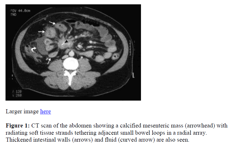
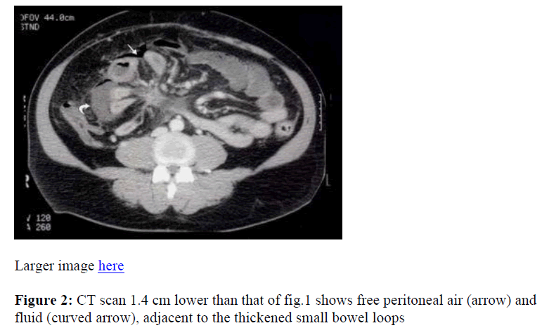
A 55-year-old male presented to the emergency room with acute abdominal pain and vomiting for more than 48 hours. The pain was diffused and nonradiating that started the same morning. The vital signs were stable. Abdominal examination revealed rigid abdomen, mild distension, guarding, and marked tenderness on palpation in the right side of the abdomen. Sluggish bowel sounds were audible. The patient had history of iron deficiency anemia with unexplained lower gastrointestinal bleeding. Laboratory investigations were unremarkable except haemoglobin of 8.4 g/dl. The patient was admitted for further evaluation. Contrast-enhanced computed tomography (CT) scan of the abdomen and pelvis was performed. Axial scans obtained with 7 mm slice thickness at 7 mm intervals with a GE helical scanner (GE medical System, Milwaukee, WI, U.S.A.). Two 8 oz cups of dilute oral contrast material (Hypaque) were administrated one hour before imaging, and 8 oz of dilute oral contrast material was given just before imaging. Approximately 125 ml of Isovue-370 contrast material (37 % iodinated; E.R. Squibb, Princeton, NJ, U.S.A.) was given intravenously. CT scan revealed a mesenteric soft tissue mass with stippled calcifications and soft tissue radiating strands that causing tethering of multiple small bowel loops (Figure 1) with localized fluid and free air adjacent to these bowel loops (Figure 2). Proximal small bowel loops were dilated. The rest of the abdominal organs are normal. Radiological diagnosis was mesenteric carcinoid tumor with small bowel perforation.
During exploratory laparotomy, the mesenteric mass was found encasing the superior mesenteric vascular supply and was nonresectable. There were necrosis and perforation of small bowel about 30 cm proximal to the ileocecal valve. A 1.5 cm mass was found proximal to the necrotic segment. The necrotic small bowel was resected. Histopathology of the resected small bowel demonstrated multiple carcinoid tumors within the bowel wall. The perforation was at the site of a focally necrotic carcinoid tumor. Postoperative course was uneventful.
Discussion
Carcinoid tumor is the most common primary neoplasm arising from enterochromaffin or enterochromaffin- like cell in the small bowel. Over 90% of carcinoid tumors are found in the gastrointestinal tract and they constitute 1.5 % of all gastrointestinal neoplasms. The most common sites of carcinoid tumor are the appendix, the terminal ileum, and the rectum [1]. Carcinoid tumors may also involve the mesentery as in this case. Lymphadenopathy and dystrophic calcification are commonly seen. Patients with carcinoid tumors may develop carcinoid syndrome, which includes flushing, intermittent diarrhea, and telanglectasia related to excessive production of serotonin. Elevation of 5-hydroxyindoleacetic acid level in the urine may be found in patients with carcinoid syndrome [4].
CT examination of this patient showed a calcified mesenteric mass with radiating soft tissue strands causing tethering of multiple loops of small bowel. Also seen was small bowel thickening and adjacent free peritoneal air and fluid that indicated small bowel neoplasia, edema, and necrosis with perforation. The mass presumably compromised blood supply to a portion of the small bowel. Proximal small bowel loops were dilated, indicating small bowel obstruction.
On CT study, a triad consisting of a soft tissue mesenteric mass, radiating linear strands and thickened adjacent bowel wall is highly suggestive of carcinoid tumor. Dystrophic calcification within the mesenteric mass is often seen [3,5]. The mesenteric mass may be a primary tumor or a metastasis from an occult or small carcinoid tumor of the small bowel. The radiating strands of the soft tissues are usually caused by tumor infiltration and fibrosis [6,7]. In this case, mesenteric vessel occlusion caused ischemia of the small bowel, leading to the necrosis and perforation, an unusual finding. An intramural neoplasm can cause small bowel obstruction. Differential diagnosis of mesenteric carcinoid includes retractile mesenteritis, lymphoma, Crohn’s disease, metastatic disease (e.g., ovary or colon), and peritoneal mestothelioma [6]. Intestinal perforation is rarely the first sign of the presence of a carcinoid as necrosis is uncommon because of its high vascularity It is important to bear this in mind while dealing with unclear pathological changes of the small intestine.
Conclusion
Perforation secondary to mesenteric carcinoid tumor is rare and its radiological findings should be kept in mind during assessment of CT scan of acute abdomen.
References
- Duvnjak M, Pavic T, Goranovic T. Perforation—a rare complication of a small- bowel carcinoid. Eur J Gastroenterol Hepatol. 2000; 12: 807-808.
- Sheth S, Horton KM, Garland MR, Fishman EK. Mesenteric neoplasms: CT appearances of primary and secondary tumors and differential diagnosis. Radio- graphics 2003; 23: 457-473.
- Pantograg-Brown L, Buetow PC, Carr NJ, Lichtenstein JE, Buck JL. Calcification and fibrosis in mesenteric carcinoid tumor: CT findings and pathologic correlation. AJR 1995; 164: 387-391.
- Buck JL, Sobin LH. Carcinoids of the gastrointestinal tract. Radiographics 1990; 10: 1081-1095.
- Cockey BM, Fishman EK, Jones B, Siegelman SS. Computed tomography of abdominal carcinoid tumor. J Comput Assist Tomogr 1986; 9: 38-42.
- Gould M, Johnson FJ. Computed tomography of abdominal carcinoid tumour.Br J Radiol 1986; 59: 881-885.
- Picus D, Glazer HS, Levitt RG, Husband JE. Computed tomography of abdominal carcinoid tumor. AJR 1984; 143: 581-584.

