ISSN: 0970-938X (Print) | 0976-1683 (Electronic)
Biomedical Research
An International Journal of Medical Sciences
- Biomedical Research (2011) Volume 22, Issue 3
Case Report: Third head of biceps brachii muscle a case study
1Dept of Anatomy, Al-Ameen medical college, Bijapur Karnataka. India.
2Department of Anatomy, Dr. V.M. Medical College, Solapur Maharashtra, India
- *Corresponding Author:
- Londhe Shashikala R.
Department of Anatomy
Al-Ameen medical college
Bijapur Karnataka
India
Accepted Date: December 17 2010
The Biceps brachii muscle is known to show variations in the number of heads. Various types of variations have been associated with the proximal attachment of Biceps brachii muscle .The Biceps brachii muscle is one of the muscle of anterior compartment of arm. It has two heads, short head and long head. This is a case report on supernumerary and vari-ant head of proximal part Biceps brachii muscle in male adult cadaver. This third head of Biceps brachii was noted unilaterally. These finding were compared with other documented reports.
Keywords
Biceps brachii muscle, third head, variation, musculocutaneous nerve.
Introduction
The Biceps brachii muscle belongs to the flexor group of muscle in arm. It is the only flexor muscle of the arm which crosses the shoulder as well as the elbow joint. As Biceps brachii muscle has two heads, short head and long head. Long head originates from the supraglenoid tubercle of scapula and short head originates from the coracoids process of scapula [1]. Distally, these two heads join to form a common tendon which inserts on the radial tube-rosity, and some aponeurotic fibers form the bicipital aponeurosis which merges with deep fascia of forearm. This muscle mainly contributes to flexion and supination of forearm [1]. The Biceps brachii muscle is innervated by the musculocutaneous nerve and supplied by brachial and anterior circumflex humeral arteries [1]. It has been reported that in 10% cases, the third head of Biceps may arise from the superomedial part of the brachialis and is attached to bicipital aponeurosis and medial side of ten-don insertion [1]. In the present case , we report a origin of third head of Biceps from upper third part of humerus at insertion of deltoid muscle in the humerus. The muscle was innervated by musculocutaneous nerve. Variations of third head of Biceps may present as a group of accessory fascicles arising from the coracoid process, the pectoralis major tendon, head of humerus, articular capsule of hu-merus or from shaft of humerus itself [2]. The presence of this accessory Biceps head may be important for aca-demic and clinical purpose. Unless symptomatic the third head of Biceps brachii may not be detected in clinical studies [3]. The third head may provide additional strength to the Biceps during supination of the forearm and elbow flexion irrespective of shoulder position [4].
The presence of third head may cause unusual bone dis-placement subsequent to fracture such variations have relevance in surgical procedure [5]. Anatomic variant that arises from persistent division between short head and long head of Biceps can be characterized with MRI know-ledge of variations in the muscle is important to character-ize injury to the components and to avoid pitfalls in im-aging diagnosis. Injuries to Biceps tendon can include tendinosis ,partial and full thickness tears and complete rupture, although injuries to Biceps tendon are less com-mon but if present that require not only correct diagnosis but also accurate characterization to appropriate guide therapy [6]. Although the third head of Biceps brachii is a common occurrence in mammal. Previous studies in hu-man being show that the third head of Biceps brachii is seen in about 8% of Chinese, 10% of Europeans, 12% of Black Africans, 18% of Japanese [7] and merely 2% in Indian population [8]. Thus the occurrence of the third head of Biceps brachii in Indian population is rare.
Case History
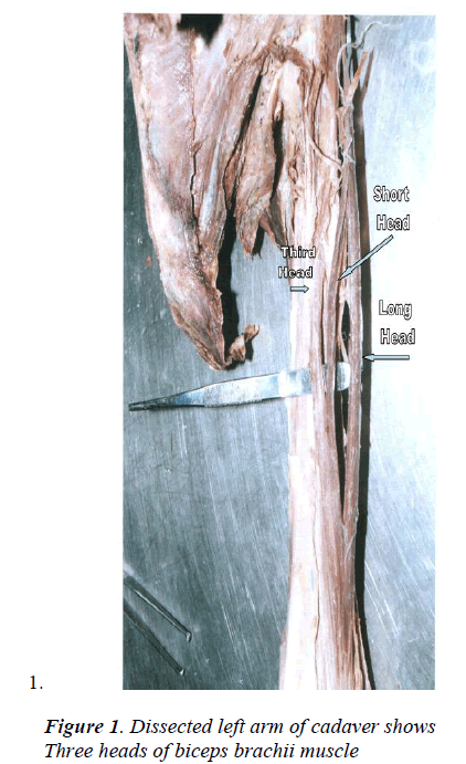
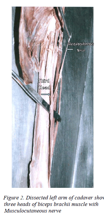
Both extremities of 10% formalin fixed 10 cadavers irre-spective of age and sex was dissected for the study. The arm was dissected carefully to display the full length of Biceps muscle from proximal attachment to the distal at-tachment. All other related structures were also exposed. The additional head was examined for the origin and course upto its insertion. Appropriate photographs were taken (Figures 1, 2). Among the 10 cadavers dissected, the third head of Biceps brachii was present in the left arm of adult male cadaver. The third head of Biceps brachii arose from upper third of humerus at the V shaped insertion of deltoid muscle (Figure 1). Third head of Bi-ceps was present medial to both long and short head of Biceps. All three heads of Biceps were supplied by musculocutaneous nerve (Figure 2). Third head descended and merged with other two heads to form common tendon and was inserted on posterior part of radial tuberosity. No other abnormalities were observed.
Discussion
Several articles have reported the incidence of third head of Biceps brachii. The Biceps brachii muscle presents a wide range of variations. They can manifest as a cluster of accessory fascicles arising from coracoid process, pector-alis minor tendon, proximal head of humerus or articular capsule of humerus [2]. Williams PL reported the inci-dence of this variation to be as much as 10%, the third head arises from the superomedial aspect of brachialis [1]. Kumar, Das, Rath [9] observed a well defined third head to arise from the anterior limb of the V shaped insertion of deltoid muscle and incidence was 3.33% [9] which is similar to our observation. Asvat et al observed that the third head of Biceps brachii originated from the humeral shaft either inferior to in common with the insertion area for the coracobrachialis or in common with the brachialis muscle. They also observed a dual origin in which medial fibres originated from the short head of Biceps and lateral fibres from the deltoid fascia. They reported an incidence of 21.5% in their study group con-sisting of blacks [10] Rai R et al stated that the occur-rence of a third head of Biceps brachii muscle is rela-tively rare in indian population. They have observed origin of third head of Biceps from anteromedial aspect of lower part of the humeral shaft. Incidence was 7.1% [11]. Kosugi et al observed that the supernumerary head of Bi-ceps arose from the humerus between the insertion of coracobrachialis and upper part of origin of brachialis and from medial intermuscular septum. They have also re-ported that in few cases, the Biceps brachii was seen to be arising from the tendon of pectoralis major , the deltoid, the articular capsule or the crest of greater tubercle of the humerus [12]. Among the reports of various stud-ies on the origin of the Biceps brachii the occurrence of supernumerary head has been the most prevalent variation within numerous reported cases of supernumerary heads of Biceps brachii muscle , the third head of Biceps bra-chii was most common. A prevalence range of 7.5% of 18.3 has been reported for the third head of biceps [12].
Kosugi et al and Asvat et al stated that there are no clear gender and racial differences in the occurrence of super-numerary head of Biceps [10,12]. Emeka, Emmanuel [13] reported unilateral third head of Biceps was lying be-tween the long and short heads. The third head was run-ning within the capsule of shoulder joint along the bicipi-tal groove to pick origin from supraglenoid tubercle of the humerus [13].
In the present case the course and branching of musculo-cutaneous nerve was normal. Hitendrakumar et al ob-served the musculocutaneous nerve to have a characteris-tic branching pattern in between the long head and the third head. Knowledge of such variation may be impor-tant for surgeons operating on the arm and clinicians di-agnosing the nerve impairment[9].
References
- Williams PL, Bannister LH, Berry MM.et al .eds.In Grays?s anatomy: The anatomical basis of medicine and surgery .38th ed. Edinburgh,ELBS Churchill Liv-ingstone 1995; PP 843.
- Sargon MF, Tuncali D, Celik HH. An unusual origin for the accessory head of biceps brachii muscle. Clin Anat 1996;9:160-162.
- Warner JJP, Paletta GA, Warren RF. Accessory head of the biceps brichii: a case report demonstrating clinical relevance. Clin Orthop Rel Res 1992; 280:179-181.
- Swieter MG, Carmichael SW. Bilateral three headed biceps brachii muscle. Anat Anz 1980; 148: 346-349.
- Vollara VR, Nagabhooshana S, Bhat SM, et al. Multi-ple accessory structures in the upper limb of a single cadaver. Singapore Med J 2008; 49 (9): e254.
- Berna Dirim, Sharon Sudarshan Brouha, Michael L. Pretterklieber, et al. Terminal Bifurcation of the Bi-ceps Brachii Muscle and Tendon: Anatomic Consid-erations and Clinical Implications. AJR 2008; 191: 248-255.
- Bergman RA, Thompson SA, Afifi AK, Saadeh FA. Compendium of Human Anatomic Variations. Balti-more: Urban & Schwarzenberg, 1988: 139-143.
- Soubhagya R, Nayak, Latha V, et al. Third head of biceps brachii: a rare occurrence in the Indian popula-tion. Ann Anat 2006; 188:159-61.
- Kumar H, Das S, Rath G. An anatomical insight in to the thirds head of biceps brachii muscle. Bratisl Lek Listy 2008; 109 (2): 76-78.
- Asvat R, Candler P, Sarmiento EE. High incidence of third head of Biceps brachii in South African popula-tion. J Anat 1993; 182: 101-104.
- Rai R Ranade AV, Prabhu LV, Pai MM, Prakash. Third head of Biceps brachii in an Indian population. Sin-gapur Med J 2007; 48 (10): 929-931
- Kosugi K, Shibata S, Yamashita H. Supernumerary head of biceps brachii and braching pattern of musculo-cutaneous nerve in Japanese. Surg Radiol Anat 1992; 14: 175-85.
- Emeka AH, Emmanuel ON. Variations of the proximal attachment of the biceps brachii muscle in a Nigerian population. International journal of anatomical varia-tions 2009; 2: 91-92.

