ISSN: 0970-938X (Print) | 0976-1683 (Electronic)
Biomedical Research
An International Journal of Medical Sciences
Research Article - Biomedical Research (2017) Volume 28, Issue 7
Comparative analysis of the medium-term effect of the medial unicompartmental arthroplasty through medial approach next to patellar and high tibial osteotomy on medial compartment osteoarthritis of the knee
1Department of Orthopaedics, School of Medicine, Jinling Hospital, Nanjing University, Nanjing, PR China
2The 66337 Troops of P.L.A, JieXiu, PR China
3Department of Counterterrorism Research, Zhejiang Police College, Hangzhou, PR China
#These authors contributed equally for this work
- *Corresponding Author:
- Zhou Xing
Department of Orthopaedics
School of Medicine, Jinling Hospital Nanjing University
PR China
Accepted date: December 28, 2016
Objective: To compare and analyse the medium-term effect of the medial unicompartmental arthroplasty through medial approach next to patellar and high tibial osteotomy on medial compartment osteoarthritis of the knee.
Methods: 72 cases of patients with medial compartment osteoarthritis of the knee were randomly divided into Group A and Group B, with 36 cases in each group. Patients in Group A were treated with medial unicompartmental arthroplasty through medial approach next to patellar. Patients in Group B were treated with high tibial osteotomy.
Results: HSS score, range of motion, VAS score, operative time and blood loss before surgery, 1, 6, 12, and 24 months after surgery in the two groups were compared. There was no significant difference of HSS score, range of motion and VAS score before surgery, 1 and 6 months after surgery between the two groups (p>0.05). The HSS score and range of motion in Group A 12 and 24 months after surgery was significantly higher than that in Group B (p<0.05). VAS score in Group A 12 and 24 months after surgery was significantly lower than that in Group B (p<0.05). There was no significant difference of operative time and blood loss between the two groups (p>0.05).
Conclusion: The near-term effect of the medial unicompartmental arthroplasty through medial approach next to patellar and high tibial osteotomy on medial compartment osteoarthritis of the knee was good. But the medium-term effect of the medial unicompartmental arthroplasty through medial approach next to patellar was more significant than the high tibial osteotomy.
Keywords
Unicompartmental arthroplasty, High tibial osteotomy, Medial compartment osteoarthritis, Medium-term effect
Introduction
Osteoarthritis of the knee, also known as degenerative arthritis, hypertrophic arthritis of the knee, is a chronic joint disease which is characterized by pathological changes such as hyperosteogeny, articular cartilage degeneration and destruction [1-4]. The clinical manifestations include morning stiffness, recurrent joint movement disorder, swelling, and pain, severe cases can cause deformity of the knee, so that the daily work and the quality of life is affected [5,6]. Unicompartmental arthroplasty and high tibial osteotomy are both effective approaches of treating medial compartment osteoarthritis of the knee [7-10]. But they are controversial in clinical. Therefore, this study was to compare and analyse the medium-term effect of the medial unicompartmental arthroplasty through medial approach next to patellar and high tibial osteotomy on medial compartment osteoarthritis of the knee, to provide some guidance in clinical.
Subjects and Methods
Object of study
72 cases of patients with medial compartment osteoarthritis of the knee treated in our hospital from May 2011 to May 2013 were selected. Inclusion criteria: a) have been audited by the hospital ethics committee; b) surgery on one knee; c) Kellgren- Lawrence grade II to III of degeneration of medial compartment articular cartilage; d) knee joint range of motion ≥ 110°, flexion contracture of<5° and varus deformity<15°; e) have signed informed consent. Exclusion criteria: a) who do not meet the above criteria; b) complicated with severe abnormalities of kidney, heart, liver, and other functions; c) with surgery contraindications; d) mental abnormalities. There were 8 cases of male and 64 cases of female, with age of 41~65, and average of (53.28 ± 7.91). The course of disease was 1~9 years, with average of (5.29 ± 1.24) years. There were 21 cases of Kellgren-Lawrence grade II and 51 cases of grade III. The patients were randomly divided into 2 groups: Group A and Group B, with 36 cases in each. There was no significant difference of general data between the two groups (p>0.05, Table 1).
| Groups | Number of cases | Male/Female | Average age (year) | Average course of disease (year) | Kellgren-Lawrence grade | |
|---|---|---|---|---|---|---|
| II | III | |||||
| A | 36 | May-31 | 52.47 ± 8.41 | 5.18 ± 1.30 | 11 | 25 |
| B | 36 | Mar-33 | 53.91 ± 7.35 | 5.40 ± 1.21 | 10 | 26 |
| X2/t | - | 0.141 | 0.774 | 0.743 | 0.067 | |
| p | - | >0.05 | >0.05 | >0.05 | >0.05 | |
Table 1: Comparison of general data between the two groups.
Methods
Group A: Patients were treated with unicompartmental arthroplasty through medial approach next to patellar. Patients were treated with routine epidural anesthesia in supine position to the medial side of the patella, and then with routine disinfection and towels. Medial incision of knee was between the upper edge of the patella and 1cm under extremity joint space, with total length of about 8 cm. At the same the joint cavity revealed out of tissues of each layer, as well as part of fat under patellar tendon was incised along the incision. Rongeur was used to remove the bone tissue with hyperplasia in edge of the medial tibiofemoral gap. The condition of the lateral articular cartilage and the integrity of the anterior cruciate ligament was checked to determine whether unicompartmental arthroplasty through medial approach next to patellar should be implemented.
Group B: Patients were treated with high tibial osteotomy. The osteotomy was carried out in the lateral proximal tibial open wedge. The upper osteotomy plane was parallel to the articular surface, retaining the medial cortex. The lower osteotomy plane was is located above the tibial tuberosity. Steel plate was used to internal fixation after osteotomy. The osteotomy angle was femoral tibial angle minus 120°, thereby ensuring 10° valgus of postoperative femorotibial anatomical axis. Supine position was used in surgery, with 90° knee bend at affected side. In which the fibula osteotomy carried out in 1/3, and the distal end of the fibula clipped about 1.0 cm lateral cortical bone. Proximal tibial osteotomy plane was about 2.0 cm under the tibial plateau, and parallel to the articular surface. The angle between distal osteotomy line and proximal osteotomy line is determined by the wedge correction degree of preoperative measurements. Typically 1.0 cm lateral tibial osteotomy can correct within 1° varus.
Analgesia, detumescence, anticoagulation and prevention of infection were carried out after surgery for both groups. The drainage tube was removed within 24 h. Patients were discharged after 10~14 days.
Observational indexes
Patients in the two groups were followed up 1, 6, 12 and 24 months after surgery. (1) Hospital for Special Surgery (HSS) knee score was sued to assess range of motion, joint function, deformity and myodynamia of knee before surgery, 1, 6, 12 and 24 months after surgery in the two groups, with scores of 85 to 100 for excellent, score of 70 to 84 for good, score of 60 to 69 for fairish, score 60 or less for poor. (2) Change of joint range of motion before surgery, 1, 6, 12 and 24 months after surgery in the two groups was observed. (3) Pain severity before surgery, 1, 6, 12 and 24 months after surgery in the two groups was observed. Standard Visual Analog Scale (VAS) was used, with score of 0 for painless, score of 1 to 3 for mild, score of 4-6 for moderate and scores of 7-10 for severe. (4) Operative time and blood loss of the two groups was observed.
Statistical analysis
SPSS19.0 software package was used for processing data, wherein for measurement data with mean ± standard deviation (x̄ ± s). Paired t-test was used for measurement data within group. Independent sample t-test was used for comparison between groups. χ2 test was used for count data. The variance analysis of repeated measurement data was used to compare the two groups of patients at different time points. p<0.05 was considered statistically significant difference.
Results
Comparison of HSS score before and after surgery between the two groups
In Table 2, there was no significant difference of HSS score before surgery, 1 and 6 months after surgery between the two groups (p>0.05). HSS score in both groups significantly increased 1, 6, 12 and 24 months after surgery (p<0.05). The HSS score in Group A 12 and 24 months after surgery was significantly higher than that in Group B (p<0.05). This indicated that the medium-term effect of knee joint of the medial unicompartmental arthroplasty through medial approach next to patellar was more significant than the high tibial osteotomy. While there was no significant difference of near-term efficacy.
| Groups | Number of cases | Before surgery | 1 month after surgery | 6 months after surgery | 12 months after surgery | 24 months after surgery |
|---|---|---|---|---|---|---|
| Group A | 36 | 54.28 ± 5.34 | 61.39 ± 7.19* | 70.28 ± 8.84* | 83.47 ± 7.63* | 93.09 ± 8.69* |
| Group B | 36 | 55.13 ± 5.01 | 60.56 ± 6.58* | 68.91 ± 7.91* | 75.15 ± 6.97* | 82.76 ± 8.13* |
| t | - | 0.697 | 0.511 | 0.693 | 4.831 | 5.208 |
| p | - | >0.05 | >0.05 | >0.05 | <0.05 | <0.05 |
| *p<0.05 compared to the same group before surgery. | ||||||
Table 2: Comparison of HSS score before and after surgery between the two groups (x̄ ± s, pints).
Comparison of joint range of motion before and after surgery between the two groups
In Table 3, there was no significant difference of joint range of motion before surgery, 1 and 6 months after surgery between the two groups (p>0.05). Joint range of motion in both groups significantly increased 1, 6, 12 and 24 months after surgery (p<0.05). The joint range of motion in Group A 12 and 24 months after surgery was significantly higher than that in Group B (p<0.05). This indicated that the medium-term effect of joint range of motion of the medial unicompartmental arthroplasty through medial approach next to patellar was more significant than the high tibial osteotomy. While there was no significant difference of near-term efficacy.
| Groups | Number of cases | Before surgery | 1 month after surgery | 6 months after surgery | 12 months after surgery | 24 months after surgery |
|---|---|---|---|---|---|---|
| Group A | 36 | 120.03 ± 1.28 | 121.96 ± 1.27* | 123.14 ± 1.03* | 126.83 ± 1.21* | 128.94 ± 1.37* |
| Group B | 36 | 120.27 ± 1.34 | 121.78 ± 1.30* | 123.13 ± 1.08* | 124.76 ± 1.38* | 126.13 ± 1.45* |
| t | - | 0.777 | 0.594 | 0.040 | 6.767 | 8.452 |
| p | - | >0.05 | >0.05 | >0.05 | <0.05 | <0.05 |
| *p<0.05 compared to the same group before surgery. | ||||||
Table 3: Comparison of joint range of motion before and after surgery between the two groups (x̄ ± s).
Comparison of VAS score before and after surgery between the two groups
In Table 4, there was no significant difference of VAS score before surgery, 1 and 6 months after surgery between the two groups (p>0.05). VAS score in both groups significantly decreased 1, 6, 12 and 24 months after surgery (p<0.05). The VAS score in Group A 12 and 24 months after surgery was significantly lower than that in Group B (p<0.05). This indicated that the medium-term effect of relieving pain in the knee of the medial unicompartmental arthroplasty through medial approach next to patellar was more significant than the high tibial osteotomy. While there was no significant difference of near-term efficacy.
| Groups | Number of cases | Before surgery | 1 month after surgery | 6 months after surgery | 12 months after surgery | 24 months after surgery |
|---|---|---|---|---|---|---|
| Group A | 36 | 7.03 ± 1.29 | 6.12 ± 0.62* | 5.03 ± 1.12* | 3.87 ± 0.59* | 2.45 ± 0.47* |
| Group B | 36 | 6.89 ± 1.23 | 6.14 ± 0.73* | 5.14 ± 0.82* | 4.59 ± 0.67* | 3.54 ± 0.50* |
| t | - | 0.472 | 0.125 | 0.476 | 4.839 | 9.530 |
| p | - | >0.05 | >0.05 | >0.05 | <0.05 | <0.050 |
| *p<0.05 compared to the same group before surgery | ||||||
Table 4: Comparison of VAS score before and after surgery between the two groups (x̄ ± s, pints).
Comparison of operative time and blood loss before and after surgery between the two groups
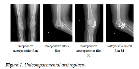
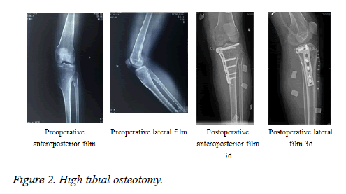
In Table 5, there was no significant difference of operative time and blood loss between the 2 groups (p>0.05). Figures 1 and 2 were the 3D weight-bearing anteroposterior and lateral X-ray film of medial compartment osteoarthritis of the knee.
| Groups | Number of cases | Operative time (min) | Blood loss (ml) |
|---|---|---|---|
| Group A | 36 | 64.82 ± 9.47 | 163.24 ± 34.10 |
| Group B | 36 | 63.47 ± 9.13 | 168.29 ± 38.74 |
| t | - | 0.616 | 0.587 |
| p | - | >0.05 | >0.05 |
Table 5: Comparison of operative time and blood loss before and after surgery between the two groups (x̄ ± s).
Discussion
Cartilage injury lesion is large, and involves two corresponding points, the tibia and femur, while the unicompartmental knee arthroplasty with high tibial osteotomy is a better choice. These two operations have been applied for decades clinically, but in terms of indications and the long-term efficacy remain controversial. Most scholars believe that high tibial osteotomy is more suitable for relatively younger patients with more activities and for patients with lesions located in the medial compartment of the varus knee. The unicompartmental arthroplasty is more suitable for relatively older and less active patients [11-14]. High tibial osteotomy includes closing wedge osteotomy and open wedge osteotomy. The main purpose of this surgical method is to correct patellofemoral joint tracks not matching caused by error line, to maintain the stability of joints and to reconstruct mechanical power lines of lower limb [15,16]. As Bonnin et al. [17] reported, 139 cases of patients treated with high tibial osteotomy were retrospectively analysed. Postoperative exercise function was significantly stronger than in the past in 20.9% of patients. 44.6% was essentially flat. The condition was slightly lower than in the past in 33% of patients. The research reports of Kyunget al. [18] indicated that high tibial osteotomy combined with arthroscopic surgery for medial osteoarthritis of the knee achieved good clinical results. Unicompartmental arthroplasty for tibiofemoral joint compartment has been for decades, but the surgical techniques, early implant design and other aspects is not perfect, therefore the effect is not very satisfactory. With the improvement of implant design and the increase of surgical experience in recent, it showed unique advantages and broad clinical applications in treatment of medial compartment osteoarthritis [19]. The main advantages of unicompartmental arthroplasty were: a) Unicompartmental arthroplasty is only for destructed mesooecium in cartilage, and does not damage normal mesooecium and cruciate ligament. b) The amount of osteotomy is small, so it preserves sufficient bone mass, which was beneficial for revision of total knee arthroplasty after surgical incident or prosthesis loosening. c) Postoperative knee flexion deformity can be corrected itself after unicompartmental arthroplasty. Parratte et al. [20] reported that unicompartmental arthroplasty treating medial compartment osteoarthritis has made good initial effect, but the long-term effects need follow-up observation. Yim et al. [21] reported that the short-term effect of unicompartmental arthroplasty combined with high tibial osteotomy on medial compartment osteoarthritis was good, but long-term follow-up observation should be carried out for long-term efficacy. The result of this study showed that HSS score and range of motion in Group A 12 and 24 months after surgery was significantly higher than that in Group B. VAS score in Group A 12 and 24 months after surgery was significantly lower than that in Group B. This indicated that the medium-term effect of the medial unicompartmental arthroplasty through medial approach next to patellar was more significant than the high tibial osteotomy, but the long-term efficacy still needed long-term follow-up.
In summary, the near-term effect of the medial unicompartmental arthroplasty through medial approach next to patellar and high tibial osteotomy on medial compartment osteoarthritis of the knee was good. But the medium-term effect of the medial unicompartmental arthroplasty through medial approach next to patellar was more significant than the high tibial osteotomy.
Funding Support
1. Military Medical Scientific Research Foundation of China (15QNP023).
2. Hospital Foundation from Jinling Hospital, Nanjing University (No.2014028).
References
- Spakova T, Rosocha J, Lacko M. Treatment of knee joint osteoarthritis with autologous platelet-rich plasma in comparison with hyaluronic acid. Am J Phys Med Rehab 2012; 91: 411-417.
- McQuade J. Osteoarthritis of the Knee. BMC Musculoskel Disord 2015; 16: 8.
- Rutjes AW, Juni P, da Costa BR, Trelle S, Nüesch E. Viscosupplementation for osteoarthritis of the knee: a systematic review and meta-analysis. Ann Intern Med 2012; 157: 180-191.
- Martin KR, Kuh D, Harris TB, Guralnik JM, Coggon D. Body mass index, occupational activity, and leisure-time physical activity: an exploration of risk factors and modifiers for knee osteoarthritis in the 1946 British birth cohort. BMC Musculoskelet Disord 2013; 14: 219.
- Wellsandt E, Gardinier E, Manal K. Association of joint moments and contact forces with early knee joint osteoarthritis after acl injury and reconstruction. Osteoarth Cartilag 2014; 22: 86-87.
- Richter M, Trzeciak T, Owecki M, Pucher A, Kaczmarczyk J. The role of adipocytokines in the pathogenesis of knee joint osteoarthritis. Int Orthop 2015; 39: 1211-1217.
- Zhai P, Sun YQ, Sun J H. Treating 46 cases of occult blood loss of primary osteoarthritis after the single knee joint replacement with tranexamic acid injection. Rheumat Arthr 2014; 14: 192-195.
- Nwachukwu BU, McCormick FM, Schairer WW. Unicompartmental knee arthroplasty versus high tibial osteotomy: United States practice patterns for the surgical treatment of unicompartmental arthritis. J Arthropl 2014; 29: 1586-1589.
- Foran JRH, Brown NM, Della Valle CJ. Long-term survivorship and failure modes of unicompartmental knee arthroplasty. Clin Orthop Relat Res 2013; 471: 102-108.
- Lee DC, Byun SJ. High tibial osteotomy. Knee Surg Relat Res 2012; 24: 61-69.
- Duivenvoorden T, Brouwer R, Baan A. Comparison of closing-wedge and opening-wedge high tibial osteotomy for medial compartment osteoarthritis of the knee. J Bone Joint Surg Am 2014; 96: 1425-1432.
- Bruyere O, Cooper C, Pavelka K, Rabenda V, Buckinx F. Changes in structure and symptoms in knee osteoarthritis and prediction of future knee replacement over 8 years. Calcif Tissue Int 2013; 93: 502-507.
- Bini S, Khatod M, Cafri G. Surgeon, implant, and patient variables may explain variability in early revision rates reported for unicompartmental arthroplasty. J Bone Joint Surg 2013; 95: 2195-2202.
- Biswas D, Van Thiel GS, Wetters NG. Medial unicompartmental knee arthroplasty in patients less than 55years old: minimum of twoyears of follow-up. J Arthroplast 2014; 29: 101-105.
- Song IH, Song EK, Seo HY. Patellofemoral alignment and anterior knee pain after closing-and opening-wedge valgus high tibial osteotomy. Arthroscop J Arthrosc Relat Surg 2012; 28: 1087-1093.
- Kim JH, Kim JR, Dong HL. Combined medial open-wedge high tibial osteotomy and modified Maquet procedure for medial compartmental osteoarthritis and patellofemoral arthritis of the knee. Eur J Orthop Surg Traumatol 2013; 23: 679-683.
- Bonnin MP, Laurent JR, Zadegan F. Can patients really participate in sport after high tibial osteotomy. Knee Surg Sports Traumatol Arthro 2013; 21: 64-73.
- Kyung BS, Kim JG, Jang KM. Are navigation systems accurate enough to predict the correction angle during high tibial osteotomy? Comparison of navigation systems with 3-dimensional computed tomography and standing radiographs. Am J Sport Med 2013; 41: 2368-2374.
- Spahn G, Hofmann GO, von Engelhardt LV. The impact of a high tibial valgus osteotomy and unicondylar medial arthroplasty on the treatment for knee osteoarthritis: a meta-analysis. Knee Surg Sport Traumatol Arthroscop 2013; 21: 96-112.
- Parratte S, Pauly V, Aubaniac JM. No long-term difference between fixed and mobile medial unicompartmental arthroplasty. Clin Orthop Relat Res 2012; 470: 61-68.
- Yim JH, Song EK, Seo HY, Kim MS, Seon JK. Comparison of high tibial osteotomy and unicompartmental knee arthroplasty at a minimum follow-up of 3 years. J Arthroplasty 2013; 28: 243-247.

