ISSN: 0970-938X (Print) | 0976-1683 (Electronic)
Biomedical Research
An International Journal of Medical Sciences
- Biomedical Research (2008) Volume 19, Issue 2
Contribution of articular and muscular structures to the limita-tion of range of motion after joint immobility: An experimental study on the rat ankle
Department of Physical Therapy, Faculty of Health Sciences, Prefectural University of Hiroshima, 1-1,Gakuen-cho, Mihara City, Hiroshima 723-0053, Japan
- *Corresponding Author:
- Sadaaki Oki
Department of Physical Therapy
Faculty of Health Sciences
Prefectural University of Hiroshima
1-1, Gakuen-cho, Mihara City
Hiroshima 723-0053
Japan
Phone/ FAX: +81-848-60-1256
E-mail: oki@pu-hiroshima.ac.jp
Accepted date: May 17 2008
To investigate contribution of articular and muscular structures to the limitation of range of motion after joint immobility, we carried out an experimental study. Unilateral ankle joints of 36 rats were immobilized in full plantar flexion with casts from the toes to below the knee joint. The rats were divided into three sets, each set having 12 rats im-mobilized for durations of 1, 2, or 4 weeks. Each set of rats was then divided into two groups (A and B), each group containing 6 rats. At the end of each period the casts were removed from 12 of the rats consecutively, and the measurements of the range of motion of the ankle joint were performed as follows. For Group A, the measurements were performed in four stages: Stage 1: before skin resection, Stage 2: after skin resection, Stage 3: after a myotomy (resection of the flexor muscles of the ankle joint), and Stage 4: after an arthrotomy (resection of capsule and ligaments of the ankle joint). For Group B, the measurements were also per-formed in four stages: Stage 1: before skin resection, Stage 2: after skin resection, Stage 3: af-ter an arthrotomy, and Stage 4: after a myotomy. There is a general tendency to presume that the muscular factor is dominant over the articu-lar factor, but there were no differences found. As a result, both the arthrogenic and the myo-genic factors contribute to joint restriction at 1, 2, and 4 weeks of immobilization.
Keywords
joint, contracture, immobilization, rat
Introduction
Joint contractures related to immobilization are a frequent problem in physical rehabilitation. Many studies have documented how joint contractures increase after immobi-lization. In these studies, histological and physiological investigations were carried out to clarify the factors re-lated to joint contractures [1-7]. Recently, it has been ex-plained that the articular structures (bone, cartilage, syno-vium/subsynovium, capsule, and ligaments) and the mus-cular structures (muscle, tendon, and fascia) are most of-ten involved in uncomplicated contractures (contractures not accompanied by neural or skin changes) [8].
For treatment, identifying which factors cause the cotrac-tures is essential. A few experiments [9,10] done to clarify the cause of the contractures involved myotomies following arthrotomies. Thus, the articular causes of the con-tracture could be observed directly. However, the muscu-lar factors of the contracture were estimated by using cal-culation [10]. In this study, our purpose was to clarify the exact effect of each factor by performing arthrotomies following myotomies, in addition to myotomies following arthrotomies. Using these procedures, each factor was directly identified.
Materials and Methods
The protocol of this study was approved by our Univer-sity Ethics Committee for Animal Experimentation. Ani-mal experiments were carried out in accordance with the National Institutes of Health Guidelines for the care and use of laboratory animals. Thirty-six female Wistar rats (10 weeks old) were used. The animals were housed in a temperature- controlled room at 20 degree C with a 12 hour light-dark cycle. The rats were provided free access to standard rat food and to water.
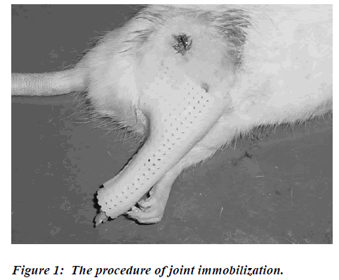
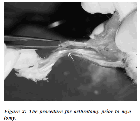
Under pentobarbital sodium anesthesia (0.5ml/kg), one ankle joint of each animal was immobilized in full plantar flexion with a cast from the toes to below the knee joint. A wire was introduced into the middle of the femur (Fig.1). The cast was anchored by a wire to prevent it from falling. The hip and knee joints of the immobilized limb were left free. The rats were randomized into three sets, each set having 12 rats immobilized for durations of 1, 2, or 4 weeks. Each set of rats was then randomized into two groups, A (n=6) and B (n=6). At the end of each immobilized period, the rats were killed with pentobarbi-tal sodium (2.0ml/kg). Their casts were removed, and, within 15 minutes, at room temperature, the measure-ments of the range of motion on the ankle joint were done. For Group A, the measurements were performed in four stages: Stage 1: before skin resection, Stage 2: after skin resection, Stage 3: after a myotomy (resection of the flexor muscles of the ankle joint), and Stage 4: after an arthrotomy (resection of capsule and ligaments of the an-kle joint). For Group B, the measurements were also per-formed in four stages: Stage 1: before skin resection, Stage 2: after skin resection, Stage 3: after an arthrotomy, and Stage 4: after a myotomy. In Stage 3 for Group B, the muscles were displaced to keep the continuity (Fig.2). After the arthrotomy was performed, the displaced muscles were returned to their original positions, and the measurements were carried out. For the measurements of the range of motion on the ankle joint, the ankle joints were kept in dorsiflexion under a 4 N-mm torque, while the knee joint was flexed maximally. Photographs of the ankle joint were taken to serve as the measurements of range of motion. Measurement of range of motion was defined as the angle (0-180 degrees) from the base line of the calcaneus to a line connecting the malleolus of the fibula and the head of the fibula.
The Wilcoxon test was used to compare the changes be-tween the four stages. Possible differences between Group A and Group B in each stage were determined by the Mann-Whitney test. All differences were considered to be significant at p<0.05.
Results
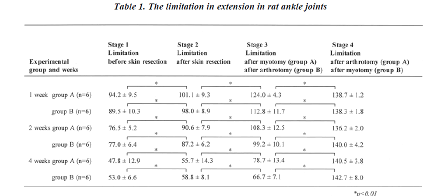
The range of motion for the ankle joints in each stage at 1, 2 and 4 weeks is summarized in Table 1. Joint restriction occurred in both groups at 1 week. The degree of the re-striction increased at 2 and 4 weeks (Stage 1). But, there were no differences between Groups A and B at 1, 2, and 4 weeks. After skin resection (Stage 2), joint restrictions decreased in both groups at 1, 2, and 4 weeks (p<0.01). In addition to Stage 2, joint restrictions decreased in Stages 3 and 4 for both groups at 1, 2, and 4 weeks (p<0.01).
The limitations of the joint were compared for Groups A and B in each stage at 1, 2, and 4 weeks. In Stage 2, there were no differences between the groups at each week. In Stage 3 of Group A, the restriction factor was the articular structure only. On the other hand, the restriction factor was muscle only in Stage 3 of Group B. In comparison between the two groups at Stage 3, the predominant cause of the contracture is different. There is a general tendency to presume that the muscular factor is dominant over the articular factor, but there were no differences found. It was found that both arthrogenic and myogenic factors contributed to the joint restriction at 1, 2, and 4 weeks of immobilization. In Stage 4, there were no differences found between both groups for each duration.
Unilateral ankle joint was immobilized in full plantar flexion with a cast from the toes to below the knee joint. A wire was introduced into the middle of the femur. The cast was prevented from falling by anchoring it with a wire.
In Stage 3 of Group B, the muscles were displaced to keep continuity (arrow). Arthrotomy was carried out in this position. After the arthrotomy, the displaced muscles were replaced into their original positions, and meas-urements were carried out under 4 N-mm torque.
Discussion
It is easy to evaluate arthrogenic restriction, as myotomy following a resection of articular structures is not a com-plicated procedure. On the other hand, to evaluate only myogenic restriction is not an easy procedure. As the ar-ticular structure is located under muscular structures, there is always a question about whether the articular structure was resected completely or not before the myotomy procedure was done. However, as we found no differences between Group A and Group B in Stage 4, it can be stated that complete resection of the articular struc-tures was achieved before the myotomy was performed. From the results of Stage 4, it is shown that our procedure is a reliable one to evaluate the factors involved in the restricted joint. As a result, we can show the distinction between arthrogenic restriction and myogenic restriction by comparing the results of Stage 3.
Trudel et al. [10] quantified the increasing role of arthro-genic changes in limiting the range of motion of joints after immobilization, especially when the period of im-mobility extended past 2 weeks. On the other hand, in our study, both arthrogenic and myogenic factors contributed to joint restriction at 1, 2, and 4 weeks of immobilization. The difference may be related to the difference in the methods. As our method provides actual results, the pre-cise causes of the contractures may be reflected in our results.
In the rat, the morphological changes in the joint that are caused by restriction of motion appear to be reversible if the period of immobilization does not exceed thirty days. After sixty days of immobilization, all major alterations consequent to the restriction of motion are permanent [1]. Intra-articular adhesions also occur after prolonged joint immobilization [1,2]. These changes are not reversible. In this study, we did not resect the intra-articular compo-nent. Actually, after the resection of the skin, muscles, capsule, and ligaments, complete dorsiflexion was achie-ved. This result shows that intra-articular adhesion does not occur within a 4-week immobilization period, as pre-viously reported.
Clinically, during the acute phase, joint exercise is widely used as the treatment for joint contractures. Joint exer-cises are effective for not just one but for both the muscu-lar and articular factors causing the contractures. How-ever, physical modalities can be applied to the specific affected areas. According to the results of our investiga-tion, it is indicated that both muscular and articular fac-tors, not just one, should be treated. The procedures that we used cannot be performed on human beings, but fur-ther investigations on experimental animals may provide more information on how treatment for the joint contrac-tures should be performed effectively.
References
- Evans EB, Eggers GWN, Butler JK, Blumel J. Experi- mental immobilization and remobilization of rat knee joints. J Bone Joint Surg Am 1960; 42: 737-758.
- Enneking WF, Horowitz M. The intra-articular effects of immobilization on the human knee. J Bone Joint Surg Am 1972; 54: 973-985.
- Tabary JC, Tabary C, Tardieu C, Tardieu G, Goldspink G. Physiological and structural changes in the cat’s so- leus muscle due to immobilization at different lengths by plaster casts. J Physiol 1972; 224: 231-244.
- Williams PE, Goldspink G. The effect of immobiliza- tion on the longitudinal growth of striated muscle fi- bers. J Anat 1973; 116: 45-55.
- Finsterbush A, Friegman B. Early changes in immobi- lized rabbits knee joints. A light and electron micro- scopic study. Clin Orthop 1973; 92: 305-319.
- Williams PE, Goldspink G. Changes in sarcomere length and physiologic properties in immobilized mus- cle. J Anat 1978; 127: 459-468.
- Williams PE, Goldspink G. Connective tissue changes in immobilized muscle. J Anat 1984; 138: 343-350.
- Trudel G. Differentiating the myogenic and arthrogenic components of joint contractures. An experimental study on the rat knee joint. Contractures secondary to immobility. Int J Rehabil Res 1997; 20: 397-404.
- Ichihashi N, Taketomi Y, Kaneko T, Yamaguchi M. Influence of skin and muscle on the knee joint stiffness in rats. Rigakuryouhougaku 1991; 18: 45-47.
- Trudel G, Unthoff HK. Contractures secondary to immobility. Is the restriction articular or muscular? An experimental longitudinal study in the rat knee. Arch Phys Med Rehabil 2000; 81: 6-13.


