ISSN: 0970-938X (Print) | 0976-1683 (Electronic)
Biomedical Research
An International Journal of Medical Sciences
Research Article - Biomedical Research (2017) Volume 28, Issue 9
Diagnostic significance of Ki67, CK19, Galectin-3 and HBME-1 expression for papillary thyroid microcarcinoma
Department of Surgical Oncology, Affiliated First People's Hospital Jiaxing Branch, Jiaxing First Hospital, Shanghai Jiao Tong University, PR China
- *Corresponding Author:
- Hong-gang Jiang
Department of Surgical Oncology
Affiliated First People's Hospital Jiaxing Branch
Jiaxing First Hospital
Shanghai Jiao Tong University, PR China
Accepted on February 1, 2017
The incidence of Papillary Thyroid Carcinoma (PTC) has increased gradually in recent years, especially for Papillary Thyroid Microcarcinoma (PTMC). The purpose of this study was to evaluate the expression of Ki67, CK19, Galectin-3 and HBME-1 protein in PTMC and evaluate the clinical significance of differential diagnosis. The specimens of 80 PTMC cases and 40 benign papillary hyperplasia thyroid cases were collected in our study. Ki67, CK19, Galectin-3 and HBME-1 protein expression were evaluated in these specimens by using immunohistochemistry.The positive expression rates of Ki67, CK19, Galectin-3 and HBME-1 in PTMC were 43%, 99%, 95% and 71%. Respectively, the rates were 15%, 38%, 25% and 15% in benign thyroid (P<0.01). Sensitivity of Ki67, CK19, Galectin-3 and HBME-1 in differential diagnosis were 43%, 99%, 95% and 71%; Specificity were 85%, 63%, 75% and 85%;Diagnostic accuracy were 57%, 87%, 88% and 76%; The area under the ROC curve were 0.638, 0.806, 0.850 and 0.781. Our study suggested that detection of Ki67, CK19, Galectin-3 and HBME-1 protein may be helpful for differential diagnosis of PTMC, and Galectin-3 was the best one in these four markers for differential diagnosis.
Keywords
Papillary thyroid microcarcinoma, Ki67, CK19, Galectin-3, HBME-1, ROC curve.
Introduction
Papillary Thyroid Microcarcinoma (PTMC) is a type of Papillary Thyroid Carcinoma (PTC), the tumor diameter of which is less than 1 cm. In the recent years, the incidence of PTC has increased gradually, especially for PTMC [1]. It is not difficult to diagnosis of PTC in general, but some smaller mass of thyroid benign lesions with papillary hyperplasia and papillary carcinoma, especially papillary microcarcinoma is difficult to distinguish, prone to misdiagnosis or missed diagnosis. Immunohistochemistry facilitate diagnosis of PTMC more easily when it is difficult to distinguish using histological criteria.
In the recent years, a large number of molecular markers in thyroid carcinoma have been used in the distinction of malignant from benign thyroid lesions. These biomarkers, such as CK19, TG, Ki67, CD56, Claudin-1, HER-2, CXCL12, TTF-1, BRAF, RET, HBME-1, Galectin-3 and so on [2]. Ki67 is a marker of cell proliferation; it has become a sensitive index of tumor cell proliferating activity. With the increasing level of Ki67 expression, the proliferative activity of tumor cells also increases [3]. CK19 is a low molecular weight cytokeratin which presents widely in simple epithelia and basal cell layers of stratified epithelium. The role of CK19 in the diagnosis of PTC is still controversial, some researcher suggested that CK19 was not a specific marker [2,4,5]. Galectin-3 is a member of oligosaccharide selective binding protein family known as lectins which plays an important role in the cell growth, cell-matrix interactions, apoptosis, neoplastic transformation and metastasis. HBME-1 is a monoclonal antibody generated against a membrane antigen of mesothelial cells. Previous researches suggested that a high positive rate of HBME-1 was observed in malignant thyroid tissues.
Immunohistochemical staining for various protein markers like CK19, galectin-3 and HBME-1 has been used frequently in the diagnosis of PTC [6-8], but the use of combinations of several proteins to evaluate the accuracy of PTMC diagnosis is still controversial. Therefore, in order to evaluate diagnostic significance of Ki67,CK19Galectin-3 and HBME-1 protein in PTMC, these four markers expression were evaluated in 120 specimens of PTMC and benign papillary hyperplasia thyroid by using immunohistochemistry in our research.
Materials and Methods
Clinical data
A total of 120 thyroid samples from the eastern part of China collected by the department of pathology in Shanghai Jiao Tong University affiliated first people's hospital Jiaxing branch between 2011 and 2013 were used in this study. There were 80 cases of PTMC, which included 13 men and 67 women, with an average age of 48 ± 12 years (range 26-76 years). All the PTMC cases underwent unilateral thyroidectomy and VI lymph node dissection. Respectively, there were 40 cases of benign papillary hyperplasia thyroid, which included 21 cases of nodular goiter, 10 cases of thyroid adenoma and 9 cases of hashimotos thyroiditis, and included 8 men and 32 women, with an average age of 51 ± 12 years (range 19-78 years). All specimens were confirmed by characteristic cytologic features and histopathology.
Reagents
The antibody included: Ki67, mouse monoclonal antibody sc-23900, 1:100, Santa cruz biotechnology, CA, USA; CK19mouse monoclonal antihuman antibody sc-53258; 1:50; Santa cruz biotechnology, CA, USA; Galectin-3, mouse monoclonal antihuman antibody clone 9C4; 1:50; Beijing Zhong Shan Biotechnology, Beijing, China; HBME-1, mouse monoclonal antihuman antibody sc-59307; 1:50; Shanghai Lan Ji Biotechnology, Shanghai, China.
Methods
All specimens were fixed in 10% neutral formalin solution, dehydrated and embedded in paraffin following conventional tissue processing, cut into 4 um sections, and stained with haematoxylin and Eosin (H&E). Immunohistochemistry (En Vision method) has been performed, according to the kit instructions for operation: (1) Paraffin sections were removed paraffin and rehydrated. (2) Paraffin sections were washed in PBS, and blocked with 3% peroxide-methanol for endogenous peroxidase ablation. (3) Sections were incubated at room temperature for 2 hours with primary monoclonal antibody. (4) Sections were washed in PBS, and incubated with biotinylated secondary antibody for 30 minutes at room temperature. (5) The reactions became visible after immersion with DAB. (6) Finally, haematoxylin staining, conventional dehydration, sealed.
Evaluation
The cells were regarded as positive for Ki67 when immunoreactivity was clearly observed in their cell nucleus. And positive for CK19, Galectin-3, HBME-1 when immunoreactivity were clearly observed in their cytoplasm or cell membrane. According to Beesley [9] classify method, immunoreactivity no staining or weak staining was considered as negative (-), staining less than 25% of the cells and buffy staining was considered as weakly positive (+), staining 25%-50% of the cells and buffy staining was considered as midrange positive (++), staining more than 50% of the cells and deep brown staining was considered as strong positive (++ +).
Statistical analysis
Data were analysed using SPSS V.17.0. The χ2 test was used for comparison of the data between the PTMC and benign papillary hyperplasia thyroid. Sensitivity, specificity and diagnostic accuracy of each marker were assessed in PTMC. The accuracy of diagnosis index was evaluated by ROC curve.
Results
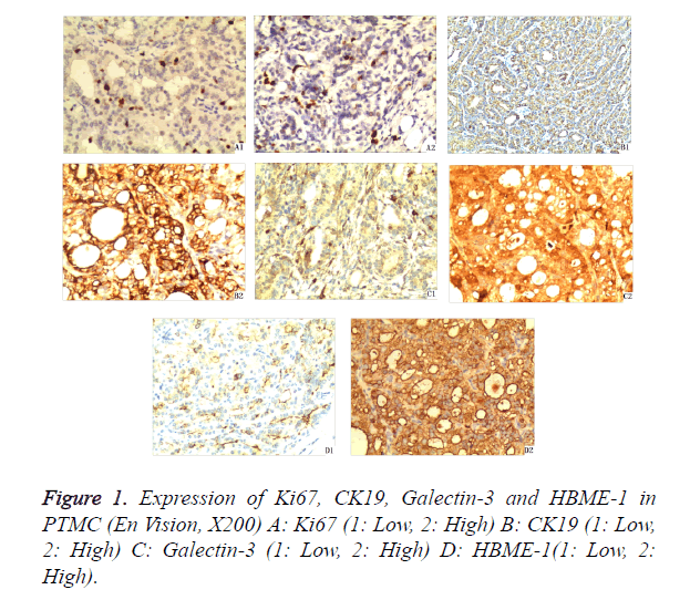
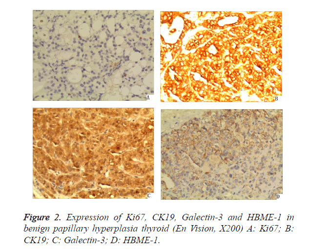
The positive expression rates of Ki67 in PTMC and benign papillary hyperplasia thyroid were 43% and 15%. There was significant difference between these two groups (χ2=9.075, P=0.003). The positive expression rates of CK19 in PTMC and benign papillary hyperplasia thyroid were 99% and 38%. There was significant difference between these two groups (χ2=58.944, P=0.000). The positive expression rates of Galectin-3 in PTMC and benign papillary hyperplasia thyroid were 95% and 25%. There was significant difference between these two groups (χ2=64.350, P=0.000). The positive expression rates of HBME-1 in PTMC and benign papillary hyperplasia thyroid were 71% and 15%. There was significant difference between these two groups (χ2=33.835, P=0.000). Details are showed in Table 1. The expression of Ki67, CK19, Galectin-3 and HBME-1 in PTMC and benign papillary hyperplasia thyroid as showed in Figures 1 and 2.
| Ki67 CK19 |
| Type cases |
| - + ++ +++ Positive rates (%) - + ++ +++ Positive rates (%) |
| PTMC 80 46 34 0 0 43 1 2 38 39 99 |
| Benign 40 34 6 0 0 15 25 15 0 0 38 |
| Galectin-3 HBME-1 |
| Type cases |
| - + ++ +++ Positive rates (%) - + ++ +++ Positive rates (%) |
| PTMC 80 4 4 36 36 95 23 2 25 30 71 |
| Benign 40 30 9 1 0 25 34 6 0 0 15 |
Table 1. Expression of Ki67, CK19, Galectin-3 and HBME-1 in PTMC and benign papillary hyperplasia thyroid.
There were one or more molecular markers positive expression in PTMC, four were positive expression of 21 cases, and three were positive expression of 49 cases. There were no positive expression cases which marked three and more than three kinds of molecules in benign papillary hyperplasia thyroid. The results of sensitivity, specificity and diagnostic accuracy of Ki67, CK19, Galectin-3 and HBME-1 in differential diagnosis as showed in Table 2.
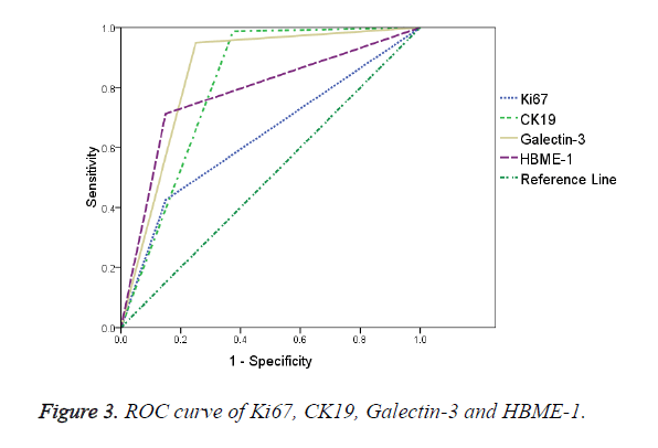
As showed in Figure 3, the area under the ROC curve of Ki67 was 0.638, the 95% confidence interval was 0.536-0.739. The area under the ROC curve of CK19 was 0.806; the 95% confidence interval was 0.710-0.903. The area under the ROC curve of Galectin-3 was 0.850; the 95% confidence interval was 0.765-0.935. The area under the ROC curve of HBME-1 was 0.781; the 95% confidence interval was 0.694-0.869.
| Sensitivity (%) | Specificity (%) | Diagnostic accuracy (%) | |
|---|---|---|---|
| Ki67 | 43 | 85 | 57 |
| CK19 | 99 | 63 | 87 |
| Galectin-3 | 95 | 75 | 88 |
| HBME-1 | 71 | 85 | 76 |
Table 2. Diagnostics performance of Ki67, CK19, Galectin-3 and HBME-1 in the distinction of PTMC from benign papillary hyperplasia thyroid.
Discussion
PTMC is a special subtype of PTC, the development trend of the young, accounting for 10.98%~28.10% of all the thyroid carcinoma cases [10]. The incidence ratio of male and female reported were different, mostly about 1:3, some as high as 1:9, the ratio was 1:5 in our study, consistent with most of the reported in the literature. The illness age was generally in 40~50 years old, the average age of this group is 48 ± 12 years old. The tumor size was small in generally, the smallest tumor length diameter was only 0.1 cm in this group. For the specimens in the case of seemingly as benign thyroid lesions, especially young women, the samples should been carefully examined, multiple sampling and especially paid attention to the calcified nodule, so as not to leak diagnosis. Although PTMC may be characterized by papillary carcinoma, such as glassy nuclei, nuclear pseudo inclusions and nuclear grooves, there were not many typical papillary thyroid carcinomas with these characteristics. And in some benign papillary hyperplasia of thyroid lesions, although there were no typical features of the papillary carcinoma, but when the nucleus increases, light staining and atypical changes, or papillary structure was not obvious and the nucleus was vacuolated, nuclear grooves, it was difficult to identify. Therefore, some auxiliary methods were needed to further clarify the diagnosis. We used immunohistochemistry to evaluate the expression of Ki67, CK19, Galectin-3 and HBME-1 protein in PTMC and benign papillary hyperplasia thyroid in our research, and analyse clinical significance of differential diagnosis.
Ki67 is a kind of DNA binding protein which is associated with cell proliferation and participates in the synthesis of DNA. It begins to appear in the G1 phase of the cell cycle, increases gradually in the S phase and G2 phase, reaches the peak in the M phase, and disappeared rapidly in the late stage of cell division [3]. There is no expression in the G0 phase [11]. At present, Ki67 has become a reliable indicator to detect the proliferation activity of tumor cell, and its positive expression indicates poor prognosis [12]. Choudhury [13] reported that strong expression of Ki67 in many malignant tumors. Actually, we have not found many data about the expression of Ki67 in PTMC. In our studythe positive expression rates of Ki67 were 43% in PTMC, and were 15% in benign. It was basically consistent with the conclusions of Aiad [14]. The sensitivity of Ki67 is low, but the specificity is relatively high in the diagnosis of PTMC. And diagnostic accuracy is not very high. We also found the absolute value of expression intensity was not very high, it may be related to the good prognosis of PTMC.
CK19 exists in a variety of epithelial and normal epithelial tumors, is strongly and diffusely expressed in papillary carcinoma, whereas it is usually absent or focally expressed in benign papillary hyperplasia of thyroid lesions [2,15]. Wu et al. [5] research suggests that in 331 cases of PTC, the positive rate of CK19 was 92.7%. Liu et al. [16] suggest that the expression of CK19 was 96.3%, for the PTC group. Respectively, for non-malignant thyroid lesions group, the expression was 40.4%. So the expression of CK19 in PTC was much higher than that in the benign thyroid lesions. In our study, the positive expression rates of CK19 was 99% in PTMC, respectively the rate was 38% in benign. The sensitivity of CK19 in the diagnosis of PTMC is very high, the specificity is relatively low, and the accuracy is high. Therefore, CK19 is a useful marker in the differential diagnosis.
Galectin-3 is a member of the lectin protein family, has a variety of biological characteristics, including cell growth regulation, cell adhesion, inflammatory reaction, cell apoptosis and so on. Galectin-3 is highly expressed in papillary thyroid carcinoma, but not in nodular goitre or adenoma. It is considered as one of the most valuable molecular markers to distinguish between benign and malignant tumors of the thyroid gland. Ma et al. [6] research suggests that in 43 cases of PTC, the positive rate of Galectin-3 was 95.3%. In our study, there was 76 cases positive expression in 80 cases of PTMC. The results of these researches were basically consistent. The positive expression rate of Galectin-3 was similar to CK19 in PTMC. The area under the ROC curve of Galectin-3 was the largest one. It suggested that Galectin-3 was better than CK19 in the differential diagnosis.
HBME-1 is a specific marker on the mesothelial cell microvilli surface. It plays an important role in the formation, growth and metastasis of tumor blood vessels. Early scholars found that it was also expressed in follicular thyroid cancer, especially in papillary thyroid carcinoma. A number of studies [16,17] found that HBME-1 was highly expressed in papillary carcinoma, but negative or partial expression in benign lesions. Liu et al. [16] suggest that the expression of HBME-1 was 85.3%, for the PTC group. Respectively, for non-malignant thyroid lesions group, the expression was 37.2%. Ma et al. [6] research suggests that in 43 cases of PTC, the positive rate of HBME-1 was 85%. In our study, the positive expression rates of HBME-1 in PTMC was 71%, respectively in benign was 15%, similar to that in previous researches. A number of studies [5,18,19] showed that HBME-1 combined with other tumor markers can be a better diagnosis of thyroid papillary carcinoma.
Leandro et al. [20] meta-analysis of 66 papers about the expression of CK19, Galectin-3 and HBME-1 in differentiated thyroid carcinoma, by immunohistochemistry, the sensitivity and specificity of CK19 were 81% and 73%, the sensitivity and specificity of Galectin-3 were 82% and 81%, the sensitivity and specificity of HBME-1 were 77% and 83%. Our study suggested that the sensitivity of CK19 and Galectin-3 were higher, but specificity were slightly lower, and the sensitivity of Ki67 and HBME-1 ware slightly lower, but the specificity was high. The area under the ROC curve of Galectin-3 was largest, so it suggested that Galectin-3 was the best of these four markers in the differential diagnosis.
Conclusion
In conclusion, when it is difficult to diagnose PTMC by morphology alone, the utilization of Ki67, CK19, Galectin-3 and HBME-1 protein combined with morphologic evaluation may be helpful to the differential diagnosis of PTMC, and Galectin-3 was the best marker in these four for differential diagnosis.
References
- Hughes DT, Haymart MR, Miller BS, Gauger PG, Doherty GM. The most commonly occurring papillary thyroid cancer in the United States is now a microcarcinoma in a patient older than 45 years. Thyroid 2011; 21: 231-236.
- Song Q, Wang D, Lou Y, Li C, Fang C. Diagnostic significance of CK19, TG, Ki67 and galectin-3 expression for papillary thyroid carcinoma in the north eastern region of China. Diagn Pathol 2011; 6: 126.
- Zhou Y, Jiang HG, Lu N, Lu BH, Chen ZH. Expression of ki67 in papillary thyroid microcarcinoma and its clinical significance. Asian Pac J Cancer Prev 2015; 16: 1605-1608.
- Chung SY, Park ES, Park SY, Song JY, Ryu HS. CXC motif ligand 12 as a novel diagnostic marker for papillary thyroid carcinoma. Head Neck 2014; 36: 1005-1012.
- Wu G, Wang J, Zhou Z. Combined staining for immunohistochemical markers in the diagnosis of papillary thyroid carcinoma: improvement in the sensitivity or specificity. J Int Med Res 2013; 41: 975-983.
- Ma H, Xu S, Yan J, Zhang C, Qin S. The value of tumor markers in the diagnosis of papillary thyroid carcinoma alone and in combination. Pol J Pathol 2014; 65: 202-209.
- Liu Z, Li X, Shi L, Maimaiti Y, Chen T. Cytokeratin 19, thyroperoxidase, HBME-1 and galectin-3 in evaluation of aggressive behavior of papillary thyroid carcinoma. Int J Clin Exp Med 2014; 7: 2304-2308.
- Das DK, Al-Waheeb SK, George SS. Contribution of immunocytochemical staining’s for galectin-3, CD44, and HBME1 to fine-needle aspiration cytology diagnosis of papillary thyroid carcinoma. Diagn Cytopathol 2014; 42: 498-505.
- Beesley MF, McLaren KM. Cytokeratin 19 and galectin-3 immunohistochemistry in the differential diagnosis of solitary thyroid nodules. Histopathology 2002; 41: 236-243.
- Zhang HX, Su C, Xie J. Clinical pathology analysis of 66 cases thyroid microcarcinoma. Chin J Cancer Prevent Treat 2010; 17: 1880-1881.
- Haroon S, Hashmi AA, Khurshid A, Kanpurwala MA, Mujtuba S. Ki67 index in breast cancer: correlation with other prognostic markers and potential in Pakistani patients. Asian Pac J Cancer Prev 2013; 14: 4353-4358.
- Klintman M, Bendahl PO, Grabau D, Lovgren K, Malmstrom P. The prognostic value of Ki67 is dependent on estrogen receptor status and histological grade in premenopausal patients with node-negative breast cancer. Mod Pathol 2010; 23: 251-259.
- Choudhury M, Singh S, Agarwal S. Diagnostic utility of Ki67 and p53 immunostaining on solitary thyroid nodule-a cytohistological and radionuclide scintigraphic study. Indian J Pathol Microbiol 2011; 54: 472-475.
- Aiad HA, Bashandy MA, Abdou AG, Zahran AA. Significance of AgNORs and ki-67 proliferative markers in differential diagnosis of thyroid lesions. Pathol Oncol Res 2013; 19: 167-175.
- Saleh HA, Jin B, Barnwell J, Alzohaili O. Utility of immunohistochemical markers in differentiating benign from malignant follicular-derived thyroid nodules. Diagn Pathol 2010; 5: 9.
- Liu Z, Yu P, Xiong Y, Zeng W, Li X. Significance of CK19, TPO, and HBME-1 expression for diagnosis of papillary thyroid carcinoma. Int J Clin Exp Med 2015; 8: 4369-4374.
- Paunovic I, Isic T, Havelka M, Tatic S, Cvejic D. Combined immunohistochemistry for thyroid peroxidase, galectin-3, CK19 and HBME-1 in differential diagnosis of thyroid tumors. APMIS 2012; 120: 368-379.
- Torregrossa L, Faviana P, Filice ME, Materazzi G, Miccoli P. CXC chemokine receptor 4 immunodetection in the follicular variant of papillary thyroid carcinoma: comparison to galectin-3 and hector battifora mesothelial cell-1. Thyroid 2010; 20: 495-504.
- Nechifor-Boila A, Borda A, Sassolas G. Immunohistochemical markers in the diagnosis of papillary thyroid carcinomas: The promising role of combined immunostaining using HBME-1 and CD56. Pathol Res Pract 2013; 209: 585-592.
- de Matos LL, Del Giglio AB, Matsubayashi CO, de Lima FM, Del GA. Expression of CK-19, galectin-3 and HBME-1 in the differentiation of thyroid lesions: systematic review and diagnostic meta-analysis. Diagn Pathol 2012; 7: 97.


