ISSN: 0970-938X (Print) | 0976-1683 (Electronic)
Biomedical Research
An International Journal of Medical Sciences
- Biomedical Research (2011) Volume 22, Issue 1
Effect of Etoricoxib on fracture healing - An experimental study
Ajit Singh1, S.K. Saraf2, R. S. Garbyal3, Shekhar4
1Department of Orthopedics, Rohilkhand Medical College, Bareilly, India
2Department of Orthopedics, Institute of Medical Sciences, Banaras Hindu University, Varanasi, India
3Department of Pathology, Institute of Medical Sciences, Banaras Hindu University, Varanasi, India
4M.P.T, Jaipur College of Physiotherapy, Jaipur, India
- Corresponding Author:
- Ajit Singh
Department of Orthopedics
Rohilkhand Medical College and Hospital
Pilibhit by pass road
Bareilly 243006, India
Accepted Date: October 27 2010
Non-steroidal anti-inflammatory drugs (NSAIDs) including selective cyclo-oxygenase-2 (COX-2) inhibitors are some of the most commonly prescribed medications worldwide. Non-selective cyclo-oxygenase (COX) inhibitors impair bone healing by inhibiting prostaglandin synthesis. The purpose of this study was to evaluate the long-term effect of etoricoxib, a new selective COX-2 inhibitor, on bone healing in rabbits, when given in therapeutic doses. Left femur was osteotomized and fixed with K wire in 36 rabbits. One group was fed etoricoxib (3mg/kg body wt/day) orally, while other group served as control. Radiological, morphological, histological and biomechanical analysis of both groups was done at 4, 8 and 12 weeks. An analysis of various parameters of study showed that etoricoxib, a selective COX-2 inhibitor, has significant inhibitory effect on bone healing at 4, 8 and 12 weeks of follow up. So it was concluded that etoricoxib significantly inhibited bone healing in rabbits especially at earlier phases of fracture healing.
Keywords
COX-2 inhibitors, Etoricoxib, Fracture healing, Prostaglandins
Introduction
Fractures and pain go side by side. Non-steroidal anti-inflammatory drugs (NSAIDs) are used widely as analgesics in the treatment of acute post-operative and traumatic pain in orthopedics. NSAIDS including selec-tive cyclo-oxygenase-2 (COX-2) inhibitors are some of the most commonly prescribed medications worldwide [1]. Non-steroidal anti-inflammatory drugs (NSAIDS) interfere with arachidonic acid metabolism by blocking prostaglandin synthesis through cyclooxygenase pathway inhibition, which has a fundamental role in bone healing [2]. Non-specific NSAIDs with both COX-1 and COX-2 inhibitory activity are known to have an adverse effect on bone healing and spinal fusion [3].
Recent trend is to use COX-2 selective NSAIDS which have been claimed to have analgesic efficacy equivalent to those of conventional NSAIDS but a lower adverse effects [4]. COX-2 selective NSAIDs were launched on basis of many potential advantages over conventional NSAIDs, such as reduced gastrointestinal bleeding and platelet abnormalities [4]. Despite widespread usage, the effects of COX-2 inhibitors on bone are not fully understood. This fact has great relevance to fracture healing, spinal fusion and Osseo-integration of prosthetic joints. The number of experimental results concerning the effects of selective COX-2 NSAIDs on bone repair is quite small, recent (from 2002 on) and also controversial [5]. In the recent past, there have been few studies showing that COX-2 selective NSAIDS inhibit long bones repair in rats and rabbits [6-9] and there are also some reports stating that they do not interfere on long bones fracture repair and on spinal fusion rates [10-13]. There is still a lot of controversy regarding the possible negative effects of cox-2 inhibitors on fracture healing .In view of these recent developments it is of major interest to establish whether or not the selective cox-2 inhibitors can be used safely as analgesics in fracture treatment. Etoricoxib is a second generation highly COX-2 selective NSAID and is the most widely used COX-2 selective NSAID. As there are no studies showing effect of etoricoxib on fracture healing, it was deemed desirable to study the effect of etoricoxib on fracture healing process in experimental animals.
Materials and Methods
White New Zealand adult healthy rabbits (n=36) with a mean weight of 2.4 kg, irrespective of sex, were obtained from animal house, Banaras Hindu University and were randomly allocated into an experimental group and a control group. Animals were kept in separate wire cage in a cycle of 12 hours light and 12 hours dark. Rabbits had free access to food and water. Etoricoxib (Nucoxia from Zydus Cadilla) was given orally to rabbits with surgically created transverse midshaft femoral fractures. Status of healing was observed clinically, radio logically, morphologically, histologically and biomechanically. The experiment was approved by the Institute Ethical Committee and is covered under section of Prevention of Cruelty to Animals Act, 1960.
Experimental design
A total of 36 rabbits were anaesthetized separately by giving intramuscular 20mg/kg of body weight of ketamine + 0.4mg/kg of midazolam + 0.5ml glycopyrollate. Two rabbits died after anesthesia and were therefore replaced by two more rabbits. Animals were placed in right lateral position on the operating table. Midshaft of left femur was exposed through a lateral incision and a transverse osteotomy of shaft of mid-femur was created using a giggly saw. Fractures were fixed by using a K-wire and wound was closed using absorbable catgut sutures. Neosporin ointment was applied and wound sealed with healex spray. All fractures were assessed to be stable by manual testing at the end of the operation. Post operatively ceftriaxone at dose of 50 mg/kg of body wt/day and amikacin at dose of 20mg/kg/day was given for 3 days and butraphenol at dose of 0.4mg/kg for pain relief was given by intramuscular route. No animal developed any sign of infection. In all cases wound healed in 5-8 days. As absorbable sutures were used, there was no need for suture removal.18 rabbits were allocated to control group and 18 to the etoricoxib treated group. Six rabbits from each group were sacrificed at 4, 8 and 12 weeks interval and were subjected to morphological, mechanical and histological examination. 18 rabbits of etoricoxib group were given the drug orally with help of disposable syringe and NG tube at daily dose of 3 mg/day/kg body weight from the day of surgery and continued till their sacrifice. The dose of etoricoxib given and the method of administration is approximately equivalent to the dose used in human beings. The animals in the placebo group were given a corresponding volume of saline at the same time points.
Radiological examination
Standard AP skiagrams of both limbs of animals were taken at 0, 4, 8 and 12 weeks. Bridging bone formation, amount of callus formation, fracture gap and fracture end sclerosis was recorded.
Morphological examination at autopsy
Animals were sacrificed by giving an overdose of ketamine and thiopentone by intramuscular route. Specimens of bilateral femur devoid of soft tissue were harvested. Medullary K-wire was removed and mobility at fracture site was tested.
Mechanical three-point bending test
This was done using INSTRON machine and a load cell of 2kg in compression mode. Three point bending test was performed under displacement control and fracture load was recorded with a load cell. Diameter and cross sectional area of callus and contra lateral femur was recorded prior to testing. The loading profile consisted of a uniform crosshead speed of 0.5mm/sec. Fracture load, bending stress; displacement and bending stiffness for both femurs in each rabbit were recorded. Values were expressed as percentage of mechanical strength of the fractured left femur compared to that of intact, contra lateral right femur. Mechanical analysis was not done in one animal in control group and two rabbits in etoricoxib group due to frank abnormality at fracture site. .
Histological study
The specimens for histological examination were fixed in 10% buffered formalin and were decalcified by keeping in 30% formic acid and then dehydrated by passing in increasing concentration of ethanol. Thereafter specimens were treated by xylene, mounted in paraffin, cut in thin sections by microtome and then cleared with xylene and alcohol before finally staining with haematoxylin and eosin. Degree of cellularity, vascularity, amount of callus, cartilage, woven and lamellar bone formation, remodeling, medullary and cortical repair etc. was observed. Rankit score [6] was used to grade the degree of fracture repair and healing in five grades. As per this grading system - Grade 0: Pseudoarthrosis formation or nonunion seen as incontrovertible cavity within cartilage plate between fracture fragments containing blood/fluid and/or lined by low cuboidal mesothelia; Grade 1: Incomplete cartilaginous union(as evidenced by retention of fibrous elements in plate); Grade 2: Complete cartilaginous union (well formed plate of hyaline cartilage uniting the fragments); Grade 3: Less than complete bony union as evidenced by presence of small amount of cartilage in fracture callus and Grade 4: Complete bony union(fracture site bridged by well formed bony trabeculae).
Statistical analysis
Calculations were done using SPSS 17 software. Results are given as mean values, and statistical significance was seen by t test for paired samples. Level of significance was set at P≤0.05.
Results
Radiological examination
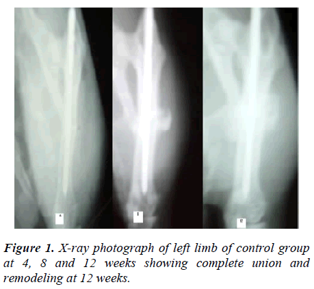
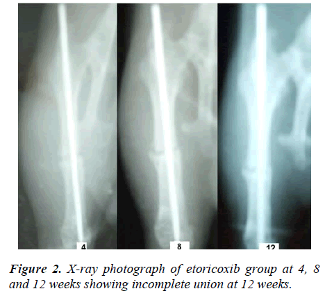
In control group, at four weeks there was mild to moderate amount of bridging callus with visible fracture line without any fracture gap seen in all rabbits. In etoricoxib group, minimal to fair amount of irregular fluffy callus was seen in all rabbits. Fracture gap was present in two rabbits. Mean amount of bridging callus was lesser than control group, although no quantitative assessment was done. At eight weeks in control group, amount of bridging callus increased in all 12 rabbits and fracture line was incompletely visible. In etoricoxib group, after eight weeks of experiment amount of bridging callus and maturity increased but callus was less mature than control group. In etoricoxib group, fracture line was visible in all rabbits, healing of fracture was poorer than controls. In control group, after 12 weeks fracture has united with variable amount of remodeling and repair of medullary canal in all six rabbits (Fig.1). After twelve weeks, in etoricoxib group variable amount of healing was seen. In two rabbits, fracture was not completely united and sclerosis of fracture ends was seen, while four rabbits had complete union. Remodeling and repair of medullary canal was poorer in etoricoxib group at 12 weeks also (Fig.2). It was inferred that fracture repair was significantly hampered by etoricoxib especially in early phases of fracture healing.
Morphological examination
All the rabbits were sacrificed at end of experiment and bilateral femur were harvested. Gross morphological changes at fracture site were observed. At four weeks in control group, there was fair to sufficient amount of immature soft callus. Frank mobility was present in one rabbit. In etoricoxib group, irregular mild to moderate amount of soft callus was seen. Quantity of callus was less as compared to controls. Frank mobility was present in two rabbits. At eight weeks, amount of callus and maturity has increased in both the groups. All the control group animals showed fair amount of mature callus and union was nearly complete. Similarly all six rabbits of etoricoxib group showed fair amount of bridging callus but the callus was Osseo-cartilaginous. No abnormal mobility was seen but the quality of callus was poorer than control group. At 12 weeks, there was a decrease in amount of callus in both groups compared with callus seen at eight weeks. All rabbits of control group showed complete bony union with mature osseous callus, while only five animals of etoricoxib group had complete bony union. One rabbit had some amount of cartilaginous union.
Mechanical analysis
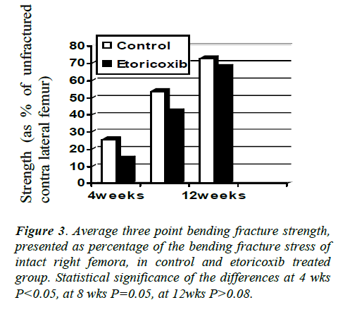
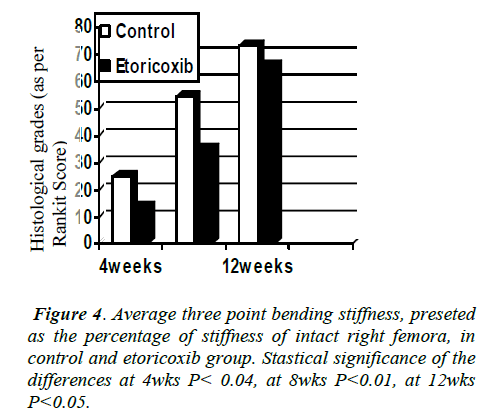
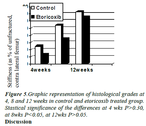
At four weeks, bending strength (P=0.039) and stiffness (P = 0.038) were significantly decreased in etoricoxib group as compared to control group. At eight weeks, etoricoxib group has significant decrease in stiffness as compared to control group (P=0.003). Although, etoricoxib group also has decreased strength compared to control group, but this difference was not significant (P=0.052). At 12 weeks, there was significant decrease in stiffness (P=0.013) in etoricoxib group as compared to control group. Although, etoricoxib group also has decreased strength compared to control group, but this difference was not significant (P=0.081). At this time specimens had approximately 70% of strength and stiffness of the contra lateral femora (Figs. 3, 4).
Histological analysis
Etoricoxib group had lower grades of histological healing at four, eight and twelve weeks, as compared to control group. Etoricoxib group showed much more fibrous tissue, lesser amount of woven bone formation and retention of some cartilaginous elements in fracture callus than control group (Fig. 5).
Discussion
The objective of the present study was to elucidate the role of etoricoxib - a COX-2 selective NSAID, in fracture healing process, with a view to understand its role in the fracture repair process. Our study shows that mechanical strength, radiographic and morphological signs of healing were significantly decreased in etoricoxib group as compared to control group at 4, 8 and 12 weeks, although differences were not very significant at 12 weeks. These results point that bone healing is impaired by COX-2 selective NSAIDS, especially in early and intermediate stages of healing. These results are in agreement with the results of previous studies on coxibs by O’Connor et al. [15], Goodman et al .[16] and Simon et al .[17] .Our study thus supports the hypothesis that COX-2 enzyme is integrally involved in the acute inflammatory response and is essential for fracture healing.
Our results are in direct contrast to the results of Gerstenfeld et al [13], Brown et al. [10] and Karachalios et al. [18] who demonstrated that COX-2 specific inhibitors did not significantly affect radiographic or biomechanical parameters of fracture healing. This difference in results is because of use of lower doses and that too for a short term in these studies. Our results also negate the inference of a recent study that etoricoxib has no deleterious effect on bone healing [19]. That study did not directly investigate the effect of a COX-2 inhibitor on bone healing but on osteoblast proliferation in vitro. Osteoblast proliferation is only one of the many processes involved in fracture healing and this probably explains difference in results.
Different COX-2 selective NSAIDS are showing different results, additional studies are required to identify a possible drug-specific bone healing response. Also most studies were done on rat or mice model and this study was done in a rabbit model, as these animals may have different pharmacodynamic and pharmacokinetic properties leading to variation in result. Another possible explanation for these variable results is that possibly there exists a baseline level of COX-2 enzyme necessary and sufficient for fracture healing and only if the inhibition is more than this baseline level, there is impairment of fracture healing.
Currently there is no established recommended dose for studies in animals. Data obtained from studies in animals cannot be used directly to interpret the effect of drugs on human beings, as there are many species-dependent differences. But despite species related differences, it would be useful if dosages used in animals were close to dosages used in humans. In our study, we selected to use a dose quite close to doses used in clinical practice. To our knowledge, this is the first experimental work on etoricoxib, in which a treatment pattern close to current recommended clinical dosage was used, and in which long-term effects of etoricoxib on bone healing were studied. Therefore future pertinent studies including an analysis of dosage dependence of effect of COX-2 specific inhibitors on fracture healing in animal models are required.
Given the potential for impaired bone healing, it is reasonable to preferentially use narcotic analgesia for pain control after fractures and avoid NSAIDS including COX-2 selective NDAIDS, if possible. Therefore we recommend about careful use of COX-2 selective NSAIDS after fractures. Though the number of animals in each group in our study was small, on the basis of findings, it can be concluded that etoricoxib (a COX-2 selective NSAID) impairs and delays the process of fracture healing, especially in early and intermediate stages. But as previous studies have showed conflicting results therefore, further analysis of effect of various COX-2 selective NSAIDS, especially etoricoxib, on fracture healing are necessary. Caution must be exercised while extrapolating these data on human beings as biological response of these drugs in humans may vary.
References
- Boursinos LA, Karachalios T, Poultsides L, Malizos KN.Do steroids, conventional non-steroidal anti-inflammatory drugs and selective Cox-2 inhibitors adversely affect fracture healing? J Musculoskelet Neuronal Interact 2009; 9 (1) :44-52.
- Li M, Thompson D, Paralkar V. Prostaglandin E2 receptors in bone formation. Int Orthop 2007; 31: 767–772.
- Abdul-Hadi O, Parvizi J, Austin MS, Eugene V, Thomas E. Nonsteroidal Anti-Inflammatory Drugs in Orthopaedics, J. Bone Joint Surg. Am 2009; 91: 2020-2027.
- Stichtenoth DO, Frolich JC. The second generation of COX-2 inhibitors: what advantages do the newest offer? Drugs 2003; 63 (1): 33-45.
- Lamano-Carvalho, Teresa Lúcia. Effect of conventional and COX-2 selective non-steroidal antiinflammatory drugs on bone healing. Acta ortop. bras 2007; 15(3): 166-168.
- Dimmen S, Nordsletten L, Madsen JE. Parecoxib and indomethacin delay early fracture healing: a study in rats. Clin Orthop Relat Res 2009; 467(8): 1992-1999.
- Gurgel BCV, Ribeiro FV, Silva MAD, Júnior N, Humberto F, Wilson SA et al. Selective COX-2 inhibitor reduces bone healing in bone defects. Braz Oral Res 2005; 19(4): 312-316.
- Endo K, Sairyo K, Komatsubara S, Takahiro S, Hiroshi E, Takayuki O et al. Cyclooxygenase-2 inhibitor delays fracture healing in rats. Acta Orthop 2005; 76 (4): 470-474.
- Leonelli SM, Goldberg BA, Safanda J, Bagwe MR, Sethuratnam S, King SJ. Effects of a cyclooxygenase-2 inhibitor (rofecoxib) on bone healing. Am J Orthop 2006; 35: 79–84 .
- Brown KM, Saunders MM, Kirsch T, Donahue HJ, Reid JS. Effect of COX-2-specific inhibition on fracture-healing in the rat femur. J Bone Joint Surg Am 2004; 86: 116-123.
- Gerstenfeld LC, Thiede M, Seibert K, Mielke C, Phippard D, Svagr B et al. Differential inhibition of fracture healing by non-selective and cyclooxygenase-2 selective non-steroidal anti-inflammatory drugs. J Orthopaedic Res 2003; 21: 670-675.
- Mullis BH, Copland ST, Weinhold PS, Miclau T, Lester GE, Bos GD. Effect of COX-2 inhibitors and non-steroidal anti-inflammatory drugs on a mouse fracture model. Injury 2006; 37(9): 827-837.
- Gerstenfeld LC, Einhorn TA . COX inhibitors and their effects on bone healing. Expert Opin Drug Saf 2004; 3: 131–136 .
- Allen HL, Wase A, Bear WT. Indomethacin and aspirin: effect of nonsteroidal anti-inflammatory agents on the rate of fracture repair in the rat. Acta Orthop Scand 1980; 51: 595-600.
- O'Connor JP, Capo JT, Tan V, Cottrell JA, Manigrasso MB, Bontempo N et al. A comparison of the effects of ibuprofen and rofecoxib on rabbit fibula osteotomy healing. Acta Orthop 2009; 80(5): 597-605.
- Goodman S, Ma T, Trindade M, Ikenoue T, Matsuura I, Wong N et al. COX-2 selective NSAID decreases bone ingrowth in vivo. J Orthop Res 2002; 20(6): 1164-1169.
- Simon AM, Manigrasso MB, O'Connor JP. Cyclo-oxygenase 2 function is essential for bone fracture healing. J Bone Mineral Res 2002; 17: 963-976
- Karachalios T, Boursinos L, Poultsides L, Khaldi L, Malizos KN. The effects of the short-term administration of low therapeutic doses of anti-COX-2 agents on the healing of fractures. An experimental study in rabbits. J Bone Joint Surg Br 2007; 89(9): 1253-1260.
- Malviya A, Kuiper JH, Makwana N, Laing P, Ashton B. The effect of newer anti-rheumatic drugs on osteogenic cell proliferation: an in-vitro study. J Orthop Surg Res 2009; 4: 17.
