ISSN: 0970-938X (Print) | 0976-1683 (Electronic)
Biomedical Research
An International Journal of Medical Sciences
Research Article - Biomedical Research (2017) Volume 28, Issue 20
Effects of oral administration of sodium nitrite on laminin expression in mice testicular interstitium: an immunohistochemical study
Sara Amini1*, Mohammad Reza Nikravesh1, Mehdi Jalali1, Alireza Fazel1 and Ariane Sadr Nabavi2
1Department of Anatomy and Cell Biology, Faculty of Medicine, Mashhad University of Medical Sciences, Mashhad, Iran
2Department of Human Genetics, Faculty of Medicine, Mashhad University of Medical Sciences, Mashhad, Iran
- *Corresponding Author:
- Sara Amini
Department of Anatomy and Cell Biology
Faculty of Medicine Mashhad University
of Medical Sciences Mashhad,Iran
Accepted Date: August 24, 2017
Background and Objective: Nitrite is one of the most common environmental contaminants affecting Extracellular Matrix (ECM) proteins expression. Drinking water contains different concentrations of nitrite and nitrate. Therefore, consumption of water contaminated with sodium nitrite can lead to infertility. This study aims to investigate the effects of oral administration of sodium nitrite on expression of laminin α5 in extracellular matrix of mice testicular interestitium using an Immunohistochemical (IHC) assessment.
Methods: Eighteen fertile male mice were divided into three groups: one control group and two treatment groups I and II. Treatment I and II groups received water containing 3 and 50 mg/L of sodium nitrite, respectively during two months. Following the sacrificing, the right and left testicle of each animal were extracted for real time PCR and immunohistochemical assessment, respectively.
Results: One-way ANOVA showed a significant difference in the laminin α5 content between the control and treatment II group. In the treatment I group, the average laminin was 0.97 fold and in the treatment II group its mean was 0.85 fold. The reduction in the amount of laminin in treatment II group was significant compared to the control group (P=0.003). The IHC tests showed that the ECM of the mice testicular interstitial tissue showed reaction to anti-laminin-α5 antibody. The intensity of the reaction in the control group and treatment I group was very weak to moderate, while the treatment II group showed weak to severe intensity. Kruskal Wallis test showed no significant difference in laminin α5 expression in the ECM of the interstitial spaces of seminiferous tubules between the groups.
Conclusion: High concentration of sodium nitrate did not change the laminin α5 expression in the ECM of the testicular interstitial tissue.
Keywords
Extracellular matrix (ECM), Immunohistochemical, Laminin, Sodium nitrite
Introduction
The first deleterious effects of sodium nitrite were revealed by observing cyanotic children. Sodium nitrite is an inorganic salt with different agricultural and industrial applications, and mankind is always subject to the resultant contaminations [1]. The treatment of animals with sodium nitrite resulted in sperm count reduction, decreased mobility, and testes weight loss. Sodium nitrite causes the irritation of evolving male germ cells by disrupting hormonal balance and tissue anoxia, and by deforming the sperms, causes the reduction of their fertility. In the biological fluids of the body, nitrate-nitrite and Nitrogen Monoxide (NO) keep transforming into one another. Nitrite and nitrate exert their biological effects by transformation into nitric oxide [2-4]. NO is reactive oxygen radical that is produced in testicular vascular endothelial cells and leydig cells, and by affecting the hypothalamicpituitarygonadal axis causes a reduction in testosterone production and inhibits steroidogenesis [5,6]. Studies have shown that there is a relationship between infertility and NO [7]. NO is located in Reactive Oxygen Species (ROS) and Reactive Nitrogen Species (RNS), which is essential for the reproductive behaviour, and if their levels exceed the natural amounts, it cause testicular dysfunction, reduction in the production of gonadotropins, and abnormality in the sperm parameters, and ultimately unsuccessful childbearing [2]. In animal studies, the drinking water impregnated with nitrite reduced the production of testosterone and testis size of the animals and made the sperm parameters abnormal and the results showed that the concentration of the nitrite/nitrate of their seminal plasma has been higher. One of the side effects of prolonged exposure to sodium nitrite is testicular toxicity [8]. Also in men who were vasectomized, the concentration of nitrite/nitrate of seminal plasma was more than other sterile men [9,10]. Laminins are glycoproteins that are involved in the organization of ECM. The composition of the ECM changes during growth and development and in response to damages and hormonal and environmental stimuli, and impact the morphology and function of the cell. Although leydig cell lacks basement membrane, but there are fragments of laminin on its surface and it synthesizes laminin-α5 subunits. ECM affects the proliferation of Leydig cell, testosterone production and gene expression, and there is a close relationship between the cell and its surrounding matrix. This relationship affects the testosterone synthesis capacity [6].
Infertility is a problem in half of married men, and environmental contaminants are among the causes of infertility in both genders. Oxidative stress, which is another cause of infertility in men, also occurs due to an imbalance between free radicals and antioxidants [11]. According to previous literature review and searching electronics resources, it seemed that the role of these contaminants in the expression of extracellular matrix proteins in the testicular tissue, which is intervenes in the behaviour of cell and tissue, has remained ambiguous, therefore, this topic became a hypothesis for proposing this project.
Materials and Methods
Eighteen fertile male mice were selected for the study. After the animals became accustomed to the new habitat, they were randomly divided into three groups of control, treatment I and treatment II. The control group enjoyed the water free of any contaminants and treatment groups I and II, received water contaminated with sodium nitrite with concentrations of 3 and 50 mg/litre, respectively, during a 60 d period. After this period, tranquil death was provided for the animals with chloroform gas inhalation and displacement of cervical spines. The left testis was first transferred to 10% formalin solution and then to 70% alcohol, for immunohistochemical technique, and their right testis was transferred to RNA later solution and was kept at -70°C.
Real-Time Technique
Total RNA extraction
Tubes containing testicular tissue were taken out of the freezer and thawed at room temperature and then according to the RNA extraction kit protocol the following steps were carried out.
First 35-40 mg of testicular tissue was removed and transferred to a 1.5 ml centrifuge tube, and using lysis buffer and needle the testis was crushed and by frequent pipetting its tissue was homogenized. Then, chloroform was added to the centrifuge tube and was vortexed and by setting the micro centrifuge on 1200 rpm and 12 min at 4°C, the tubes were centrifuged, and the upper phase of the tube was transferred to a new centrifuge tube. At the next stage, 70% ethanol was added to the tube and was spun, the mixture was transferred to the spin column and was centrifuged for one minute at 13000 rpm, and its sediment was drained and by adding PW to the columns, it was centrifuged for another minute at 13000 rpm.
The centrifuge tubes were spun for two minutes at 13000 rpm until the remaining PW was discharged from the environment, and the columns were transferred to a new 1.5 ml centrifuge tube. 40 μl DEPC-treated water was added to each centrifuge tube and was spun for another minute at 13000 round, until pure RNA was obtained and it was kept at -70°C for the next step [11].
cDNA synthesis
Following the protocol of the kit of the manufacturing company of the cDNA synthesis Easy Kit, the steps of cDNA synthesis were carried out. First, in each centrifuge tube, 5 μl total RNA, 2 μl Oligo-dt and 1 μl Radom hexamer were mixed together, and by adding DEPC-treated water, the volume of the solution was brought up to 10 μl. The above mixture was incubated in thermocycler at 65°C/5 min. Then, 10 μl RT Premix was added to the solution in order for its volume to be brought to 20 μl, and the solution was pipetted several times. In the following, it was incubated for ten min at 25°C and for 60 min at 50°C, and after being incubated for ten minutes at 70°C, the reaction was terminated [12,13].
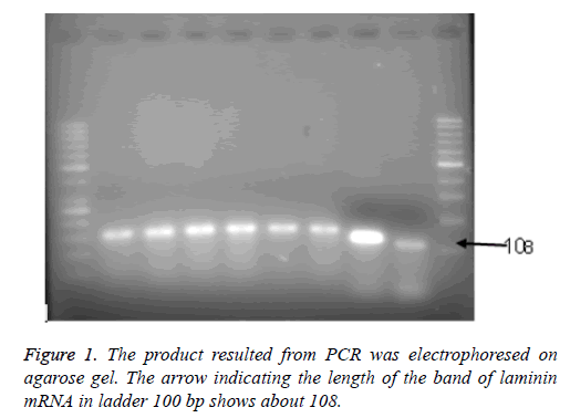
In order to ensure the results, 5 μl of the PCR (Polymerase Chain Reaction) product was electrophoresed on 2% agarose gel, which contained 0.5 mg/litre of ethidium bromide, and was analyzed using gel documentation system (Figure 1).
Primer design
To determine the expression of mRNA of laminin α5 in testicular tissue, four primers were designed using Designer Bacon, as shown in Table 1. The sequence of each primer was obtained from NCBI web site, and then, exon and intron regions of the mRNA sequences of each gene were aligned, and using BLAST software the Primer binding specificity of each gene was ensured. To prevent the proliferation of gene fragment on the DNA chain, an intron was placed between the primers (Table 1) [12,13].
| Primer | Gene sequences | |
|---|---|---|
| Laminin-α5 | Forward | 5-CGTCCCACAGGAATAGGCT-3 |
| Reverse | 5-TACCAACGAAGGGCTGCG-3 | |
| Beta-actin | Forward | 5-GGGAAATCGTGCGTGACA-3 |
| Reverse | 5-TCAGGAGGAGCAATGATCTTG-3 |
Table 1. The specifications of laminin and beta-actin primers.
Preparation of primers
The primers were the product of the Korean Macrogen company. According to the protocols of the abovementioned company, for the preparation of a concentration of 10 pmol of each primer, a proper volume of distilled water was added to each tube containing the forward and reverse primers until a 100 solution of each one of them was achieved [11].
RT-PCR technique
Before starting the RT-PCR, gel electrophoresis was performed to ensure accurate operation of the primers and the length of their bands was obtained (Figure 1). No primer dimer was formed during the amplification cycles. The thermal curve program of the Applied Biosystems Step One machine was set and the main solution of the process was procured, and the amount of laminin was measured semi-quantitatively using SYBR Green.
Changes in the laminin α5 expression in each group was compared with housekeeping Beta-actin gene, and ΔΔCT model was used to calculate it, and the results were presented in the form of fold change, compared to the control group. The ΔCT of the gene is determined by subtracting the CT value of the laminin gene from the CT value of the β-actin gene. ΔΔCT of the control sample is zero, thus -20=1. Therefore, the fold change compared to the control group was one [14].
Immunohistochemical technique
Before performing this technique, the routine procedures of tissue processing were performed and the prepared five micron slices were transferred to polyllysine slides, and then, the specific procedures of the IHC technique were performed, which we will discuss briefly.
After the clearing of tissues using xylene, the slides were passed through decreasing concentrations of alcohol in order for the tissues to be hydrated. Then, in order to prevent internal peroxidases activity, the slides were incubated in 3% hydrogen peroxide solution (diluted in methanol), and after that they were washed with distilled water, and according to antigen retrieval heat method, the slides (immersed in buffer) were placed for ten minutes in warm water bath at 100°C. After their cooling at room temperature and washing them with distilled water, they were placed in Tris buffer containing Triton X100 with a concentration of 0.25 for five minutes and they were also placed in buffer (containing 10% goat serum and 1% bovine serum albumin), during the night the slides were exposed to laminin antibody (with a ratio of 1:150), at 4°C temperature, and after washing with buffer containing Triton X100 (0.025) they were exposed to secondary antibody (conjugated to HRP) with a ratio of 1:800, at room temperature for two hours. Then, they were exposed to PBS containing 3, 3'-Diaminobenzidine and hydrogen peroxide. After three washes in running water and distilled water, in order to establish the background color, they were immersed in hematoxylin for a few seconds. Next, they were again washed with running water and distilled water and then they were passed through 90% alcohol, absolute alcohol, and three xylene containers and then they were mounted. The slides were evaluated using an Olympus optical microscope, and according to Table 2, the ranking of the intensity of the developed brown color was performed [11].
| Reaction intensity | Without reaction | Very weak | Weak | Moderate | Severe |
|---|---|---|---|---|---|
| Degrees | 0 | 1 | 2 | 3 | 4 |
Table 2. The ranking criterion for the intensity of the reaction based on the developed color on tissue slice sections from without reaction to dark brown.
Statistical analysis
SPSS software was used for data analysis. Nonparametric Kolmogorov-Smirnov test was used to evaluate the normality of the data distribution, and one-way ANOVA was used to compare mean quantitative factors. Since the data distribution was normal, to compare the qualitative variables in the groups, Kruskal-Wallis test was used, and their dual comparison was performed with the MannWhitney test.
The results were presented in the form of (Mean ± SEM), and P<0.05 was considered significant (Table 2).
Results
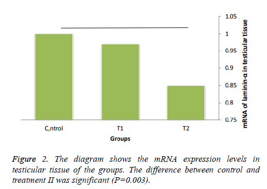
According to one-way ANOVA, there was a significant difference in the value of laminin α5 between the control and treatment II groups. In the 3 mg/l treatment group, the amount of the presence of laminin in animals was 1.1, 1.1, 0.9, 0.9, 0.95 and 0.9 and with the mean of 0.97 and in the 50 mg/l treatment group the level of presence in testicular tissue was 0.9, 0.85, 0.8, 0.9, 0.9, and 0.8, which its mean was 0.85. The reduction in the level of laminin in treatment II was significant with respect to the control group (P=0.003), but the difference with treatment I was not significant (Figure 2).
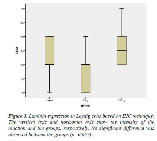
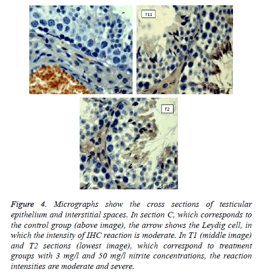
The ECM of the testicular interstitial tissue of the mouse showed reaction to anti-laminin-α5 antibody. This reaction in the animals of the control group was in the range of very weak to moderate (with weak median), and in the animals of treatment I group that received the concentration of 3 mg/l of sodium nitrite, it was in the range of very weak to moderate (weak median), and in the group with 50 mg/l concentration of sodium nitrite, it was in the range of weak to severe (weak and average median). There was no significant difference among groups (p=0.057, (Figures 3 and 4)).
Figure 4: Micrographs show the cross sections of testicular epithelium and interstitial spaces. In section C, which corresponds to the control group (above image), the arrow shows the Leydig cell, in which the intensity of IHC reaction is moderate. In T1 (middle image) and T2 sections (lowest image), which correspond to treatment groups with 3 mg/l and 50 mg/l nitrite concentrations, the reaction intensities are moderate and severe.
Discussion and Conclusion
The results of this study showed that the oral intake of sodium nitrite at a dose of 50 mg/l does not cause serious changes in laminin α5 expression in interstitial ECM of testis.
In our previous study, consumption of contaminated drinking water with excessive levels of sodium nitrite (50 mg/l), caused an increase in laminin α5 expression in the ECM of the apical compartment of the seminiferous epithelium, and it might affect the behaviour of cells by disrupting the composition of ECM [14,15]. However, in the present study, we observed no changes in the expression of this protein in testicular interspatial spaces indicating that spermatogenic germ cells might be more sensitive to sodium nitrite, compared with the leydig cells.
Nitrite is a more stable metabolite of NO that is produced endogenously. Leydig cell produces a small amount of NO, which not only inhibits the synthesis of testosterone, but the NO donors also inhibit steroidogenesis in Leydig cells. On the other hand, the NO synthase inhibitors increase testosterone.Reduction in the synthesis of testosterone, which is triggered by the shock of the animal immobilization is mediated through the NO path [2,4,16]. In the experimental model for inflammation of the testis, the number of macrophages in the testicular interstitial spaces increased, which was associated with the degeneration of germ cells and disruption of steroidogenesis. Therefore, Dietrich et al. concluded that NONOS system plays a role in the damage to testicular function by the inflammation, and NO, which is produced mainly from macrophages, causes damage to Leydig cells by inducing oxidative stress [17].
In this study we observed no change in the expression of laminin α5 in interstitial spaces. Moreover, no histological change was observed in these cells, such as pyknosis and cellular architecture. Therefore, it seems that sodium nitrite in this study, has not caused the destabilization of NO concentration, and therefore, has not compromised the leydig cell function.
Laminin α5 is among proteins that are expressed in the testis tissue of mice. The ECM has a direct role in organizing cell metagenesis, cell proliferation, migration, morphology, and restoration [18]. In the cultivation environment, Leydig cells have loose connection with collagen type I and V, but they have a strong connection with fibronectin, laminin, collagen type IV, and basement membrane components. Leydig cells are adjacent to the capillaries and venules, which themselves are surrounded by a network of lymphatic sinusoids. Collagen type IV and laminin have a direct relationship with Leydig cells. These proteins are found on the surfaces of Leydig cell and also between adjacent Leydig cells. The extracellular matrix affects the cell structure and function [19]. Therefore, any changes in the expression of proteins can affect the morphology, cell adhesion, cell cohesion, integrity of interstitial cells, and also settlement of seminiferous tubules [5]. Although in the present study, the cell ultrastructure and the study were not performed using electron microscopy, but since there was no tangible change in the expression of laminin, it is unlikely that sodium nitrite can impair the function of the cells of interstitial spaces.
Leydig cells are settled by attaching to the basement membrane of the blood and lymph peripheral vessels, and any changes in the environmental matrices can affect steroidogenic capacity of Leydig cells and their steroidogenesis [19]. In the present study, with regard to the fact that sodium nitrite has not caused any increase or decrease in laminin α5 expression in interstitial spaces, it can be said that it has not changed the composition of the matrix and therefore, has not caused the malfunction of the cells of this region, including Leydig cells.
Leydig cells express sGC (soluble guanylyl cyclase) and NOS (NO synthase) enzymes in their cytoplasm that one of them is a producer of NO and the other one is the mediator of its effects. There is no consensus on the effect of NO on testosterone production. In one hand, this radical has inhibitory effects on testosterone production in vivo, and on the other hand, it has stimulatory effects on testosterone production in vitro. Furthermore, some researchers concluded that the NO associated inhibitory effects on steroidogenesis depend on its concentration where low concentrations result in stimulatory effect and high concentrations result in inhibitory effects [20].
In the present study, which was carried out relying on immunohistochemistry and real-time techniques, but the concentration of enzymes and oxidants were not measured, nonetheless we could say that in this study it is also possible that nitrite to have caused the inhibition of nitrite and/or the reduction of superoxide dismutase (SOD) enzyme activity, and by the disruption of the balance between oxidants and antioxidants in favour of oxidants, to have upset the testicular tissue, but given the lack of change in laminin α5 expression, it appears the Leydig cell to have remained unaffected or at least through this protein no damage to have been inflicted to the cells present in the interstitial spaces.
In the study performed by Dietrich et al., the role of NO/NOS in the development of testicular inflammation was investigated. Orchitis was induced in the animals. Cellular infiltration was associated with fibrosis, testicular atrophy and infertility. Also, Leydig cell hyperplasia and hypertrophy associated with increased intratesticular testosterone were observed. Increased infiltration of macrophages in testis was associated with increased production of NO, which has an important role in cell mass conversation and steroidogenesis. They concluded that the NO/NOS system has a role in suffering the testicular function in orchitis, and the NO secreted by macrophages can affect the function of leydig cells [21,22]. In this study, sodium nitrite did not cause any change in histological characteristics of the leydig cells, and the cells remained intact. Therefore, we presume that oral administration of sodium nitrite did not cause a detectable change in the concentration of NO, and the residing cells in the testicular interstitial spaces were not upset.
The present study is the first effort that examines the role of nitrite and nitrate in testicular extracellular matrix proteins expression. In the previous paper we reported that sodium nitrite concentrations exceeding the allowable limits had caused changes in laminin α5 expression in apical compartment of the seminiferous epithelium tubules. In this study, high concentrations of nitrite, even though significantly reduced the total amount of the laminin in testicular tissue, but did not create any change in its expression in the extracellular matrix of the testicular interstitial tissue, therefore, it seems that leydig cells had been resistant to sodium nitrite. The authors of this paper believe that more detailed studies, with higher dosages, and with non-oral administration are required in order for the impact of this contaminant to be clarified on the ECM proteins expression in testicular interstitial spaces.
Acknowledgment
This manuscript is a part of PhD thesis of Mashhad University of Medical Science in Iran and supported by the research Grant No. 930439.
The authors wish to thank Mrs. Motejadded and Mrs. Tajik for cooperation in research. We also want to thank the staff of the Animal Facilities of Mashhad University of Medical Sciences.
References
- Fewtrell L. Drinking-water nitrates, methemoglobinemia and global burden of disease: a discussion. Environ Health Perspect 2004: 1371-1374.
- Doshi SB, Khullar K,Sharma RK, Agarwal A. Role of reactive nitrogen species in male infertility. Reprod Biol Endocrinol 2012; 10: 109.
- Pant N, Srivastava S. Testicular and spermatotoxic effect of nitrate in mice. Hum Exp Toxicol 2002; 21: 37-41.
- Boink A, Dormans J, Speijers G, Vleeming W. Effects of nitrates and nitrites in experimental animals. Special Publication-Royal Society of Chemistry 1999; 237: 317-326.
- Guerriero G, Trocchia S, Abdel-Gawad FK, Ciarcia G. Roles of reactive oxygen species in the spermatogenesis regulation. Front Endocrinol 2007; 141.
- Mazaud Guittot S, Vérot A, Odet F, Chauvin MA, le Magueresse-Battistoni B. A comprehensive survey of the laminins and collagens type IV expressed in mouse Leydig cells and their regulation by LH/hCG. Reprod 2008; 135: 479-488.
- Loveland K, Schlatt S, Sasaki T, Chu M, Timpl R, Dziadek M. Developmental changes in the basement membrane of the normal and hypothyroid postnatal rat testis: segmental localization of fibulin-2 and fibronectin. Biol Reprod 1998; 58: 1123-1130.
- Pavolva E, Dimova D, Petrova E, Glucheva Y, Atanassova N. Changes in rat testis and sperm count after acute treatment with sodium nitrite. Bulg J Agricult Sci 2013; 19.
- Skinner M, Tung P, Fritz I. Co-operativity between Sertoli cells and testicular peritubular cells in the production and deposition of extracellular matrix components. J Cell Biol 1985; 100: 1941-1947.
- Battaglia C, Giulini S, Regnani G, Di Girolamo R, Paganelli S, Facchinetti F. Seminal plasma nitrite/nitrate and intra-testicular Doppler flow in fertile and infertile subjects. Hum Reprod 2000; 15: 2554-2558.
- Sara A, Mohammad Reza N, Mehdi J, Alireza F, Ariane SN. Effects of sodium nitrite on fibronectin expression of the testicular parenchyma in mice. Int J Adv Biotechnol Res 2017.
- Kohbanani MS, Nikravesh MR,Jalali M, Fazel A, Sankian M, Bideskan AE. Effects of maternal nicotine exposure on expression of laminin alph5 in lung tissue of new-born. Pakistan J Biol Sci 2012; 15: 1168.
- Pahang H, Nikravesh MR, Jalali M, Bideskan AE, Zargari P, Sadr nabavi A. Effects of maternal nicotine exposure on expression of laminin alpha 5 in lung tissue of newborn. Pakistan J Biol Sci 2012; 15: 1168.
- Pffafi MW. A new mathematical model for relative quantification in real-time RT-PCR. Nucleic Acids Res 2001; 29: 45.
- Sarah A, Mohammad Reza N, Mehdi J, Alireza F, Ariane Sadr N. Evaluation of the laminin α5 expression in mice testicular parenchyma following an exposure to water contaminated with sodium nitrite. Int J Adv Biotech Res 2016; 7: 513-522.
- Tatjana K, Silvana A, Radmila KE, Desanka M. The involvement of nitric oxide in stress-impaired testicular steroidogenesis. Euro J Pharmacol 1998; 267-273.
- David KP, Vaclav P. Nitric oxide is a mediator of the inhibitory effect of activated macrophages on production of androgen by the leydig cell of the mouse. Endocrinol 1998; 139: 922-931.
- Ismo V, Jouni L, Taneli T, Matti K, Robert EB, Veli-Pekka LA, Limo L. Distinct changes in the laminin composition of basement membranes in human seminiferous tubules during development and degeneration. Am J Pathol 1997; 150: 4.
- Dnduchis B, Scheteingart H, Cigorraga S, Vianello SE, Casanova MB, Lustig L. Immunodetection of cell adhesion molecules and extracellular matrix proteins in rat Leydig cell cultures. Int J Androl 1996; 19: 353-361.
- Valenti S, Cuttica CM, Fazzuoli L, Giordano G, Giusti M. Biphasic effect of nitric oxide on testosterone and cyclic GMP production by purified rat Leydig cells cultured in vitro. Int J Androl 1999; 22: 336-341.
- Sabrina JD, Mónica IF, Patricia VJ, Cristian MAS, Livia L, María ST. Inhibition of NOS-NO system prevents autoimmune orchitis development in rats: relevance of no released by testicular macrophages in germ cell apoptosis and testosterone secretion. PLoS One 2015; 10.
- Gayta´n F, Bellido C, Aguilar R, Morales C, van Rooijen N, Aguilar E. Role of the testis in the response of the pituitary-testicular axis to nitric oxide-related agents. Euro J Endocrinol 1997; 137: 301-308.


