ISSN: 0970-938X (Print) | 0976-1683 (Electronic)
Biomedical Research
An International Journal of Medical Sciences
- Biomedical Research (2014) Volume 25, Issue 2
Evaluation of Holter Results in Patients with Obesity: A Cross Section Study.
1Izmir Tepecik Training and Research Hospital, Family Practice Clinic, Turkey
2Izmir Tepecik Training and Research Hospital, Cardiology Clinic, Turkey
3Izmir Katip Çelebi University, Faculty of Medicine, Department of Family Medicine, Turkey
- *Corresponding Author:
- Kurtuluş ONGEL
Department Head of Family Medicine
Izmir Katip Çelebi University, Izmir, Turkey
Accepted date: January 23 2014
This study aims to investigate the changes at heart rate and cardiac rhythm of obese patients admitted to obesity polyclinic using rhythm holter electrocardiography (ECG). Following the ethics committee approval, 114 patients admitted to Izmir Tepecik Training and Research Hospital Obesity Polyclinic between November 2012 and February 2013 were randomly assigned and monitored by rhythm holter device. The values of average heart rate (AHR), maximum heart rate (MAXHR), minimum heart rate (MINHR) and time domain parameters as SDNN, SDANN and RMSSD were recorded and their relation with obesity parameters and laboratory findings were analyzed. While 105 (92,10%) of 114 patients were female, 9 (7,90%) of them were male and the average age was 45.82±11.39 (min:16; max:69). The AHR, MAXHR, MINHR, SDNN, SDANN and RMSSD of them were 78.01±8.41 (min:56; max:109), 136.28±17.69 (min:105; max:181), 51.64±7.11 (min:24; max:67), 140.06±44.40 (min:47; max:368,45), 151.71±87.55 (min:15.13; max:415.00), and 65.55±52.83 (min:20.85; max:402.67), respectively. Statistically significant relation was found between the age and AHR, MAXHR and SDANN (p<0.01). Significant relation in regard to waist circumference was detected with only AHR and SDNN (p<0.05). Similarly, the only significant relation in regard to body mass index was detected with MAXHR (p:0.01). A decrease in heart rate variability is associated with the risk of coronary artery disease. Heart rate variability declines along with enlarging waist circumference. In the follow up process of obese patients, waist circumference monitoring comes out to be substantial rather than the overall body weight monitoring.
Keywords
Holter, obesity, rhythm
Introduction
As one of the modern world’s diseases, obesity is a cause of disproportionate increase at body fat tissues. According to World Health Organization’s (WHO) data, there are approximately 300 million obese people throughout the world and approximately 50% of the whole population are expected to be obese by 2025 [1]. Metabolic syndrome, which asserts itself with intraabdominal obesity, dyslipidemia, increased blood pressure, insulin resistance and/or glucose intolerance associates obesity [2,3].
Obesity is detected according to the body mass index (BMI) and waist circumference (WC) values. BMI more than 30 kg/m2 and WC over 94 cm and 80 cm respectively for males and females are considered as obesity. Obesity is classified as phase 1 (BMI: 30-40), phase 2 (BMI: 40-50) and phase 3 (BMI: >50).
Waist circumference gives more advise than BMI about the body fat distribution. At the same time, it is related with cardiovascular disease risk since it is a visceral adiposity measurement. The fat tissue cells accumulated in visceral adiposity are different than subcutaneous fat tissues. Visceral adipositis are both larger and functionally active. They synthesize leptin, TNF alpha, angiotensinogen and convert cortisone to cortisol. They involve adrenergic receptors. Hence, sympathetic system activation increases at obesity [4]. It is known that excessively active sympathetic nervous system; causing diabetes type 2, increased heart rate, vascular resistance and sodium retention, triggers cardiovascular diseases [5]. At the same time, visceral adiposity increase arises with an increase at the insulin secretion from pancreas due to the impact of changes at parasympathetic system. Consequently, metabolic syndrome comes out [5,6]. The changes at the autonomous system together with the hormonal changes could also lead to arrhythmia at obese patients.
Together with 10% weight increase, there will be a decrease at the parasympathetic system which is accompanied by a heart rate increase [7,8]. However, during the weight reduction process, heart rate decreases [9]. Such changes arising at vagal activities as a result of weight increase might be a mechanism of arrhythmia and other cardiac anomalies accompanying to obesity. These results lead up to the explanation of obesity as a major and modifiable risk factor for heart disease [10,11].
Obesity increases the risk of sudden death due to cardiac dysfunction and arrhythmia [12]. Myocardial adiposity and dilated cardiomyopathy at morbid obese people may cause fatal arrhythmia. It is observed at the study of Framingham that weight increase leads to an increase at the risk of sudden death at female and male [12-15].
The heart rate variability is a parameter in the evaluation of the function of autonomic nervous system. Decreased heart rate variability is a risk factor for cardiovascular mortality and morbidity. In this respect, it is a method that can be used confidentially also at obese patients as in case of many other clinical conditions [16,17]. The aim of this study is to analyze the relation of rhythm holter ECG parameters with obesity diagnosis criteria.
Materials and Methods
Following the ethics committee approval, 114 of the patients applied to Izmir Tepecik Training and Research Hospital Obesity Polyclinic between the dates November 2012 and February 2013, were randomly assigned. Monitoring the patients for 24 hours by Holter-ECG, time and frequency domain heart rate variability analysis was held. The values of average heart rate (AHR), maximum heart rate (MAXHR), minimum heart rate (MINHR) and time domain parameters such as SDNN, SDANN, RMSSD were recorded and relations of these values with obesity parameters and laboratory findings were analyzed.
The patients using drugs that may affect heart rate variability (digital, beta blocker and calcium channel blockers not covered by dihydropyridine group) and patients who has heart failure constituted the exclusion criteria.
Digital holter monitor was used for 24 hours based Holter-ECG records. While the parasites were cleaned semi manually during the analysis, the time domain KHD analysis was made automatically. SDNN, SDANN and RMSSD were taken as the time domain parameters [18]. The descriptions of time domain measurements of heart rate variability are submitted at Table 1.
Results
While 105 (92,10%) of the 114 patients included to the study were female, 9 (7,90%) of them were male and the average age was 45,82±11,39 (min:16-max:69). While the body mass index was within the range of 25-30 for 13 (11,40%) patients, it was between 30-40 for 74 (64,91%) patients and above 40 for 27 (23,69%) patients. Average waist circumference (WC) was found as 110,94±9,52 (min:87-max:137) cm, whereas average body weight was calculated as 91,16+13,4 (min:66-max:127) kg. When the electrocardiograms of patients were evaluated; 2 bradycardia, 1 branch block and 1 extrasystole was observed. The biochemical parameters of the patients are summarized at Table 2.
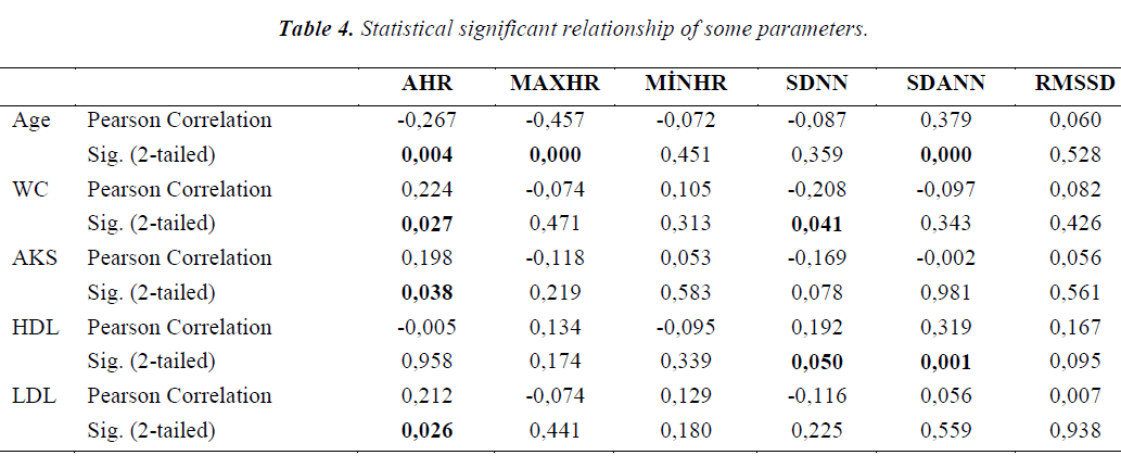
Time domain rhythm holter parameters of patients are summarized at Table 3 Statistically significant relationship couldn’t be detected between the patients’ weight, body mass index and heart rate variability parameters. When the correlation between the obesity parameters and laboratory values with rhythm holter findings has been analyzed, statistically significant relationship was detected between age and AHR, MAXHR and SDANN (respectively p<0.01). On the other hand, there was a significant relationship between WC and AHR and SDNN (p<0.05). While there was no significant statistical relationship between the patients’ body weights and cardiac values, MAXHR was the only one detected as having a significant relationship with BMI (p<0.01). Statistically significant relationship between biochemical parameters and SDNN and SDANN couldn’t be detected (p>0.05). Statistical significant relationship of some parameters of the patients are summarized at Table 4.
Fifteen of the 114 patients (13,1%) were sent with verbal recommendations about their diet regime. 1400 kcal. diet for 27 patients (23,7%), 1600 kcal. diet for 42 patients (36,9%) and 1800 kcal. diet for 30 patients (26,3%) were prescribed. After 6 months follow-up; average weight loss was 5,5±0,3 (min:0,1-max:23) kg
Discussion
Excessive weight or obesity is a complex and multifactorial chronic disease. It progresses as a result of the interaction between the genotype and environment. It constitutes a base for many diseases. One of these impacts is the reduction at heart rate.
Heart rate variability gives advise regarding the response of heart to the autonomic signals that originates as a consequence of sympathetic and parasympathetic balance. Therefore, Pieper SJ and his friends underline the importance of heart rate variability analysis in the context of the evaluation of cardiovascular response to autonomous tonus changes [19].
Heart rate variability indicates the body autonomic activity change and is accepted as its reflection to cardiovascular system. The reduction at heart rate variability particularly indicates the risk of increased arrhythmia after myocardial infarction (MI) and heart insufficiency and it is always pathological. Evaluation of patients in this direction is done by rhythm holter ECG examination. Heart rate variability is evaluated with the data collected by holter monitorization at 24 hours bases.
SDNN sets out the neurocardiac influence and is a confidential parameter of heart rate variability. SDNN value less than hundred (<100) - which means its reduction- points out the risk of coronary artery disease [19]. Although the average SDNN value of the patients included to the study was calculated as 140+44.4, SDNN values of 15 patients were found as less than 100. The body mass index of the patients having SDNN values less than 100 was scanned. The lack of a relationship between BMI and SDNN and the existence of a significant relationship between waist circumference and SDNN assert the importance of abdominal obesity. In the study, it is underlined that heart rate variability decreases due to the increase at waist circumference rather than body mass index. This gives the impression that particularly waist circumference is more important in regard to cardiac follow up at obese patients. The study held by Karason and his friends also claims that obesity leads up to a reduction at heart rate variability and the increased sympathetic activity and decreased vagal activity are recovered after weight loss. These researchers also observed the reduction at blood pressure and norepinephrine secretion together with weight loss [20].
Increase at average heart rate and parasympathetic system activity along with 10% weight increase was shown at the study of Hirsch J. and his friends. As a result of this study; researchers mentioned that parasympathetic system may be effective in weight increase and thus it may lead to arrhythmia or different cardiac problems at obese patients [9]. Mentioned result also supports this study.
It is highlighted that the importance of heart rate variability measurements -which is a non invasive method- in making diagnosis, risk identification and follow up will increase. Additionally, the cardiovascular complications of diseases creating secondary autonomic function disorder such as obesity are also accepted in this group [21]. Similarly, it is stated at another study held in 1995 that reduced parasympathetic system activity due to increased body weight is associated with the increased cardiovascular risk and scales up the frequency of sudden death [22].
As a result, the primary follow up of waist circumference is important in the evaluation of obese patients since enlarged waist circumference creates a cardiac risk. Monitoring waist circumference more regular than body weight at obesity clinics is more significant in detecting the risky individuals in terms of coronary artery disease.
References
- World Health Organization 2008. Global strategy on diet physical activity and health. http://www.who.int/dietphysicalactivity/en/ Accessed on: 10.08.2013
- Aktuglu MB, Yılmaz M, Alioglu T, Karaali Z, Velet M, Yigit N, Tunca O, Sayılan S, Keskin I, Kendir M. Evaluation of treadmill stress test results of patients with their pre and post test hsCRP and NT-proBNP levels. Smyrna Tıp Dergisi 2012; 2: 15-21.
- Ford ES, Giles WH, Dietz WH. Prevalence of the metabolic syndrome among US adults: findings from the third National Health and Nutrition Examination Survey JAMA 2002; 2873: 356-359.
- Bahceci M. Obesity. In: TEMD Obesity, Dyslipidemia. Hypertension Study Group eds. Turkish Association of Endocrinology and Metabolism. Hypertension, Obesity and Lipid Metabolism Diagnosis and Treatment Guide. İstanbul, 2011; 50-80.
- Perin PC, Maule S, Quadri R. Sympathetic nervous system, diabetes and hypertension. Clin Exp Hypertens 2001; 23: 45-55.
- Kreier F, Fliers E, Voshol PJ, Van Eden CG, Havekes LM, Kalsbeek A, Van Heijningen CL, Sluiter AA, Mettenleiter TC, Romijn JA, Sauerwein HP, Buijs RM. Selective parasympathetic innervation of subcutaneous and intra-abdominal fat: functional implications. J Clin Invest 2002; 110: 1243-1250.
- Després JP. Is visceral obesity the cause of the metabolic syndrome? Ann Med 2006; 38: 52-63.
- Reis JP, Macera CA, Araneta MR, Lindsay SP, Marshall SJ, Wingard DL. Comparison of overall obesity and body fat distribution in predicting risk of mortality. Obesity 2009; 17: 1232-1239.
- Hirsch J, Leibel RL, Mackintosh R, Aguirre A. Heart rate variability as a measure of autonomic function during weight change in humans. Am J Physiol 1991; 261: 1418-1423.
- Eckel RH. Obesity and heart disease: A statement for healthcare professionals from the Nutrition committee, American Heart Association. Circulation 1997; 96: 3248-3250.
- Eckel RH, Krauss RM. American Heart Association call to action: Obesity as a major risk factor coronary heart disease: AHA Nutrition Committee. Circulation 1998; 97: 2099-2100.
- Kannel WB, Plehn JF, Cupples LA. Cardiac failure and sudden death in the Framingham Study. Am Heart 1998; 115: 869-875.
- Messerli FH, Sundgaard-Rise K, Reisin ED, et al. Dimorphic cardiac adaptation to obesity and arterial hypertension. Ann Intern Med 1983; 99: 757-761.
- Clinical Guidelines on the Identification, Evaluation and Treatment of Overweight and Obesity in Adults-The Evidence Report: National Institutes of Health. Obes Res 1998; (Sppl: 2) 51S-209S.
- Bouchard C, Després J, Mauriége P. Genetic and Nongenetic determinants of Regional fat distribution. Endocr Rev 1993; 14: 72-93.
- Sztajzel J, Golay A, Makoundou V, Lehmann TN, Barthassat V, Sievert K, et al. Impact of body fat mass extent on cardiac autonomic alterations in women. Eur J Clin Invest 2009; 39: 649-656.
- Thayer JF, Yamamoto SS, Brosschot JF. The relationship of autonomic imbalance, heart rate variability and cardiovascular disease risk factors. Int J Cardiol 2010; 141:122-131.
- Malpas SC, Maling TJ. Heart-rate variability and cardiac autonomic function in diabetes. Diabetes 990; 39: 1177-1181.
- Corr PB, Yamada KA, Witkowski FX. Mechanisms controlling cardiac autonomic function and their relation to arrhythmogenesis. In: Fozzard HA, Haber F
- Jennings RB, Katz AN, Morgan HE, eds. The Heart and Cardiovascular System. Newyork, Raven Pres, 1986:1343-1403.
- cKarason K, Molgaard H, Wikstrand J, Sjostrom L. Heart rate variability in obesity and the effect of weight loss. Am J Cardiol 1999; 83: 1242-1247.
- Kayıkcıoglu M, Payzın S. Heart variability. Turkish Cardiology Association Archive 2001; 29: 238-245.
- Petretta M, Bonaduce D, De Fılıppo E, Mureddu GF, Scalfı L, Marcıano F, Bıancı V, Salemme L, De Sımone G, Contaldo F. Assessment of cardiac autonomic control by heart period variability in patients with early onset familial obesity. Eur J Clin Invest 1995; 25: 826-832.


