ISSN: 0970-938X (Print) | 0976-1683 (Electronic)
Biomedical Research
An International Journal of Medical Sciences
Research Article - Biomedical Research (2017) Artificial Intelligent Techniques for Bio Medical Signal Processing: Edition-I
In the teaching of oral and maxillofacial medical imaging with CBCT technology
1Department of Stomatology, Renji Hospital, School of Medicine, Shanghai Jiao Tong University, 145 Middle Shandong Road, Shanghai, PR China
2Department of Orthodontics, Ninth Peoples Hospital, Shanghai Jiao Tong University School of Medicine, Shanghai Key Laboratory of Stomatology, Shanghai, PR China
- *Corresponding Author:
- Jiameng Yu
Department of Stomatology
School of Medicine
Shanghai Jiao Tong University, PR China
Accepted on January 14, 2017
Compared to the traditional CT technology, CBCT technology has more advantages. The application of CBCT technology in the teaching of oral and maxillofacial medical imaging is less in our country, which was not conducive to the understanding of operation of students in clinical practice as well as the relevant theoretical knowledge. Firstly, the working principle and system composition of CBCT system were described in order to better understand its application in Teaching, which showed the advantages compared to the traditional CT Technology. The learning results after completing the theoretical knowledge of traditional CT technology and CBCT technology and the corresponding clinical practice were studied according to packet control tests of medical students in a university in 2016. The conclusion was obtained that CBCT has a positive effect on the teaching of oral and maxillofacial medical imaging, which not only can make students master the operation of the related instruments and equipment, but also can make it more accurate for reading and comparative analysis so as to learn the pathological characteristics of the patient and helpful for clinical diagnosis. Therefore, the application of CBCT technology in the teaching of oral and maxillofacial medical imaging should be strengthened.
Keywords
CBCT technology, Oral and maxillofacial medicine, Imaging, Teaching.
Introduction
The use of X-ray CT technology was widely used in the daily medical diagnosis and treatment process. It was be able to scan the human body by X-ray imaging technology. And the image is reconstructed according to the different attenuation of the image. The pathological characteristics of the patients was analyzed according to the image [1]. CT technology can not only directly determine the patient's physical condition, but also in the operation is very convenient and quick. CBCT technology was successfully developed and gradually applied in clinical practice at the end of last century. Although this technology has been used in some hospitals in our country at present, it has not been widely popularized [2]. Compared with the traditional CT technology, the CBCT system not only has better imaging effect, but also has a higher utilization rate of X-rays. The time required is relatively small and the acquisition speed is faster. CBCT is a reliable in assessing the proximity of vital structures which may interfere with orthodontic treatment. It is much more reliable and accurate than the traditional methods. It can be predicted that the general application of CBCT technology is the trend of the hospital. Although the CBCT technology has been involved in the teaching of oral and maxillofacial medical imaging in China, it was not explained in depth [3]. A group of medical students in a university were divided into two groups in order to explore the application of CBCT technology in the teaching of oral and maxillofacial medical imaging. The two groups of students respectively studied on the traditional CT technology and CBCT technology theoretical knowledge and conducted the clinical practice of two different systems in hospital. Finally, the two groups of students’ study situation were conducted statistical analysis so as to understand the impact of CBCT technology on students on the oral and maxillofacial medical imaging teaching effect.
CBCT Technology
CBCT system working principle
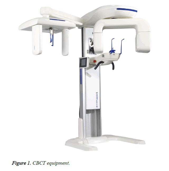
CBCT (Figure 1) is called Cone beam CT. CBCT imaging technology is a cone beam CT imaging technology, whose working principle is the same as the traditional CT technology, which is the use of X-ray projection to the object after the material absorption and attenuation of the nature [4]. The radiation that is not absorbed will continue to propagate along the original path when the X-rays penetrate the object because there is a difference between the thickness and the density of the penetrated object. So X-ray is absorbed by the situation is not the same and there are differences in the remaining energy [5]. The remaining X-ray energy is measured by the detector for photoelectric conversion and generates projection data. Complete the imaging within 360 degree range finally. Form a three-dimensional reconstruction image by setting the CT reconstruction algorithm [6].
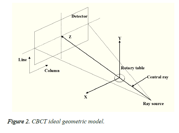
The CT reconstruction algorithm includes two kinds of analytic reconstruction algorithm and iterative reconstruction algorithm mainly. The former is fast, but its anti-noise ability is poor. Although the latter has a strong anti-noise ability, it needs a large amount of data. As a result, the reconstruct time is long and it is less used in clinical practice [7]. CBCT system is commonly used in the FDK algorithm. That is the use of fan beam filtered back projection algorithm for three-dimensional expansion and achieves image reconstruction ultimately. The typical set model of CBCT technology is shown in Figure 2.
CBCT system composition
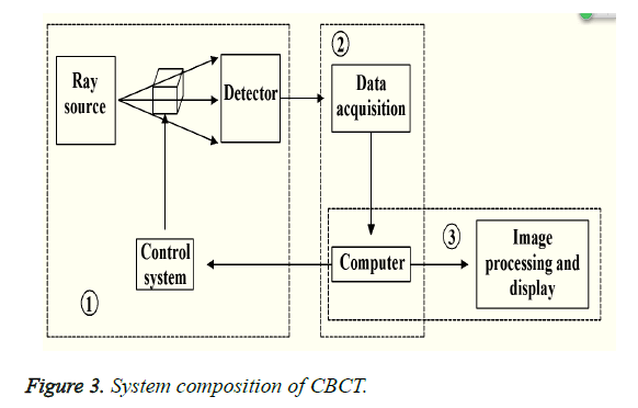
The CBCT system mainly has three parts: the control and the scanning part, the data collection part and the image processing and the display part. These three parts form an organic whole. The whole process of scanning, collecting data and imaging was completed [8]. The scanning system includes three parts: rotary table, X-ray source and detector. Data acquisition system is to transfer the collected data to the computer and then complete the storage. Image processing system is to store the information in accordance with the set of calculation method for image reconstruction, and display on the screen for the user to provide the image observation, analysis, comparison [9]. Its system composition is shown in Figure 3. In Figure 3 the labels 1, 2, 3 respectively represent the CBCT system in the scanning system, data collection system, image processing system.
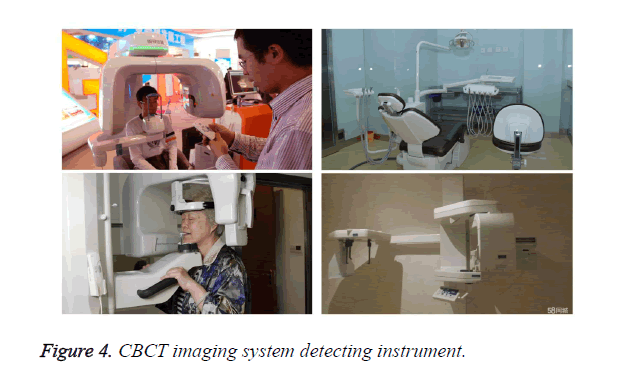
Nowadays, CBCT system was widely used in various medical specialties. The application of oral and maxillofacial medicine is more successful, because the system is able to carry out three-dimensional reconstruction of bone structure. CBCT has a considerable advantage in the field of maxillofacial bone structure, periodontal tissue, implant and other structures compared with the previous CT imaging system [9]. It can be directly or indirectly quantified in the measurement of the bone defect. The alveolar bone defect can be measured and the space is more images. Compared with the traditional CT imaging system, the advantages of CBCT are as follows: (1) The time required for scanning is short because of the shorter exposure time. Therefore, it is necessary to carry out a rotation to obtain a three-dimensional image, thus avoiding the impact of distortion and overlap [10]. (2) It requires only one as described in mind, which makes the random error reduction. (3) The images obtained can include images of sagittal, coronal and axial planes. Therefore, it is helpful for doctors to analyze and study the maxillofacial region more accurately in practice [11]. (4) The software function of CBCT system is more perfect and the calculation accuracy is higher. The computer can even select analysis method for numerical analysis automatically. It can be more convenient to directly show the situation of the jaw face (Figure 4) [12]. (5) It can be rotated from various angles to carry out research and observation because it is three-dimensional modeling and imaging, and even according to the data for image reconstruction [13].
The Teaching Research of CBCT Technology in the Oral and Maxillofacial Medical Imaging
Research method
Imaging knowledge and oral medicine are related in various professional including the oral implant, oral and maxillofacial surgery, periodontal disease, which is a basic course that every oral medicine professional doctors have to master (Table 1) [14]. Nowadays, CBCT technology has been more and more used in various hospitals and related medical structures, but it has not been widely used. Many hospitals are still using the traditional CT technology. Failing to excavate and utilize the advantages of CBCT technology is not conducive to the development and progress of Medical Science in China, which increases the diagnostic rate of misdiagnosis of some medical diagnosis and affects the health of the people of our country in a certain extent [15].
| School name | Class hours of theory course | Class hours of experimental class | Total hours | |
|---|---|---|---|---|
| Abroad | Dallas Texas Dental College | 1258 | 3758 | 2016 |
| Glasgow University Medical College | 1190 | 2265 | 3455 | |
| School of Dentistry, University of Liverpool | 1188 | 2118 | 3306 | |
| Domestic | School of Dentistry, University of Hong Kong | 1699 | 2623 | 4322 |
| College of dental medicine, Capital Medical University | 2224 | 3276 | 5500 | |
Table 1. Comparison of teaching at home and abroad.
Although CBCT technology has been widely used in the teaching of oral and maxillofacial medical imaging, the teaching content is not rich enough and teaching methods are not mature enough as well as the lack of adequate clinical practice [16]. Therefore, you should allow students to practice by way of clinical practice in the completion of adequate theoretical teaching and master the theoretical knowledge in-depth through the practice. Medical students in a university in 2016 were divided into two groups to conduct control experiment in order to study the difference of the application effect of traditional CT technology and CBCT technology in the teaching of oral medicine. The two groups of students are the same level students. One group of the theoretical knowledge was conducted traditional CT technology teaching, which was known as the traditional teaching group. Another group of CBCT technology was conducted theory of knowledge teaching, which was known as the control group. In addition the difference in the CT technology professors to the two groups of students, the rest of the medical professional knowledge is consistent. Traditional teaching group have a total of 65 people and 67 people in the control group. Two groups of students were in the relevant on knowledge base.
Two groups of different teaching groups selected a few local hospitals for practice after the completion of the theoretical knowledge of the professor. Traditional teaching group adopted the traditional CT system and the control group used the hospital CBCT system (the performance of CBCT system is shown in Table 2). After 14 days of practice, the students were graded according to the situation of the students. Compare the learning situation according to the scores situation of the two groups of students. The students return to school and the teacher organized to carry out the test of theoretical knowledge after the completion of the internship. Finally, the feedback information of the students was carried out statistical investigation and learning effect under different CT Technology Teaching was conducted comprehensive examination.
| X- ray beam | Vertebral body |
|---|---|
| Focus | 0.5mm (IEC336) |
| Sensor technology | CMOS sensor and CCD- optical fiber sensor |
| Gray level | 16374-14bit |
| Minimum spatial pixel size | 76 × 76 × 76 μm |
| Scan time | 4-16 seconds |
| Reconstruction time | Within 2 minutes |
| Imaging field of vision | 50 × 37 mm |
Table 2. Instrument technical parameters.
Research process
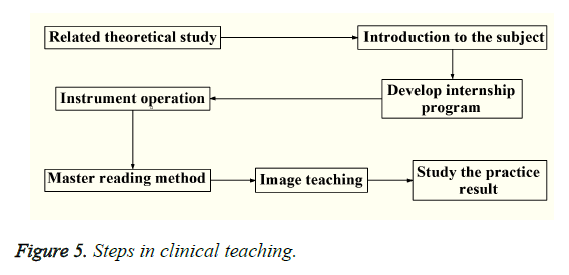
The traditional teaching group and the control group have completed their respective theoretical knowledge and started to practice in several local hospitals on April 20, 2016. Enter the hospital department of oral medicine in the practice process firstly. Teachers introduce the Department of the situation to students, focusing on the introduction of oral and maxillofacial medical imaging related content. Secondly, the students develops internship program. Arrange according to the clinical task of the Department of radiology of the College of oral medicine after the teacher view. Give students the actual operational opportunities as far as possible to in ensuring the normal operation of the Department. The specific teaching steps in the hospital are as in Figure 5.
The first step should be to carry out the operation of the instrument exercises and familiar with all kinds of imaging equipment in the course of the specific practice of the hospital. It can be cast by these machines after mastering the use of various types of equipment. The projection method for oral and maxillofacial is more in medicine including Fahrenheit, oral panoramic root splinters etc. Therefore, the method that students have to master is more and it needs the teacher to carry out a full teaching guidance. Demonstrate the students' reading analysis method after the completion of the skills of the cast. Let it be able to have a basic judgment of the quality of the photos and analysis of the image in the film to identify the changes in the rational. You can observe the location of the lesion, size, density, shape, as well as the relationship with the surrounding tissue, etc when conduct analysis of these films. The patient's condition has a preliminary judgment combined with other clinical examination results. It is very important to the patient's clinical diagnosis and should to make use of the image data to accurately judge. It requires doctors to have sufficient theoretical knowledge and reading experience. We can analyze compare and analyze according to the images with different pathological changes. We should grasp the different images which reflect the same pathology and image reflects as well as characteristics of different pathology in particular [17].
It should to allow students to fully grasp the operational capabilities of the instrument, reading ability, comparative analysis ability, autonomous learning ability and the ability to access information through the practice of the hospital. The ability of the students to cultivate the effect also have a greater difference because of there is a big difference between the actual clinical application in the traditional CT technology and CBCT technology. For example, the imaging effect of the CBCT system is better and clearer, and the result is more accurate. Students can better understand the pathological characteristics of the patient using CBCT technology and get more information through the image. Teachers access to students’ five ability to master after 14 days of practice. The total score of the individual ability is 10 points, and the average score of the two groups of students is calculated. Observe the ability of students to cultivate and enhance the situation according to the scores of two different techniques.
Results and Discussion and Analysis
The traditional and the control group were taught using the images of various patients. The control group were taught using panoramic and CT scans whereas the control group used only CBCT imaging. The CBCT examinations were carried only after the history and clinical examination of the patient was done. The lowest radiation dose through which adequate resolution for diagnosis is possible was used in CBCT.
Results study
The teacher made an assessment and grade for all the students after 14 days of clinical practice. The score reflects the ability of the students. The score results are as in Table 3.
| Entry name | Traditional teaching group | Control teaching group | P |
|---|---|---|---|
| Operating capability (10 points) | 7.52 ± 0.61 | 8.61 ± 0.59 | <0.01 |
| Reading ability (10 points) | 6.91 ± 0.49 | 8.02 ± 0.55 | <0.01 |
| Comparative analysis (10 points) | 7.71 ± 0.70 | 8.84 ± 0.72 | <0.01 |
| Autonomous learning ability (10 points) | 6.83 ± 0.47 | 8.11 ± 0.53 | <0.01 |
| Information acquisition capability (10 points) | 6.55 ± 0.51 | 8.07 ± 0.48 | <0.01 |
Table 3. Practice assessment score.
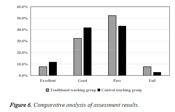
We can see through the score results that the ability to operate, read pieces, contrast analysis ability, self-learning ability and information acquisition capability of score of the students using CBCT technology in clinical practice is higher than traditional teaching group. The imaging result of CBCT technology in the actual clinical practice is better than the traditional CT technology in previous studies. Therefore, the students learning CBCT technology grasp the ability to read and the compare and analyze more easily in the internship. Judge the patient's pathology through the film. The student's study enthusiasm is higher because of the more accurate judgment. All the students return to school and the teacher develops the examination questions as well as conduct unified theoretical knowledge of the test after the completion of the teaching process. Assessment results were evaluated for evaluation in accordance with the outstanding, good, pass, fail the four different criteria (Table 4). The proportion of secondary school students in accordance with the four different standards is analyzed in order to make the analysis more accurate because the number of the two groups is not exactly the same (Figure 6).
| Team | Traditional teaching group | Control teaching group | ||
|---|---|---|---|---|
| Number | Proportion | Number | Proportion | |
| Excellent | 5 | 7.7% | 8 | 11.9% |
| Good | 21 | 32.3% | 28 | 41.8% |
| Pass | 34 | 52.3% | 29 | 43.3% |
| Fail | 5 | 7.7% | 2 | 3.0% |
Table 4. Comparison of assessment results.
As can be seen from the figure above, the traditional teaching group of students’ theoretical knowledge test scores is poorer to the theoretical knowledge of the control group. The results were excellent in 11.9%, good accounting for 41.8% in the control group. The outstanding achievement is 7.7%, and good accounting for 32.3% in traditional teaching group. As a result, the students have more in-depth knowledge of the corresponding theoretical by using of CBCT technology for the relevant teaching. Conduct to the control group of students in the questionnaire survey with a total of 67 people. The main content of the survey is to understand the impact of CBCT technology on its learning. The survey results are as in Table 5.
| Project | Control teaching group | |
|---|---|---|
| Number | Proportion | |
| Helps to master the instrument operation | 61 | 91.0% |
| It is helpful in the clinical diagnosis | 59 | 88.1% |
| Helps to stimulate interest in learning | 61 | 91.0% |
| Improve the learning efficiency and efficiency | 63 | 94.0% |
| It is more applicable to the teaching of image | 60 | 89.6% |
Table 5. Student feedback.
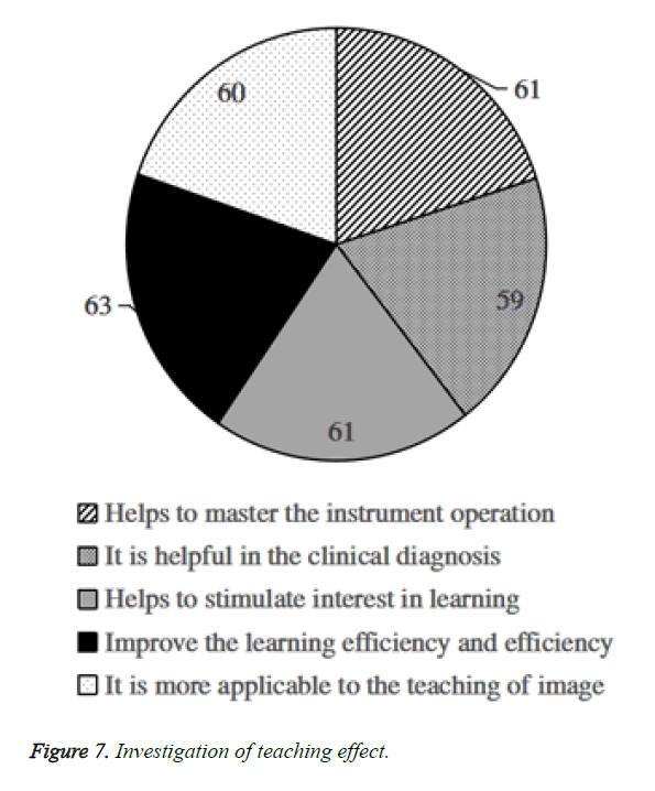
We can see through the student's feedback information that the use of CBCT technology in the impact of teaching helps students to master the instrument operation and carry out clinical diagnosis, which can stimulate students' interest in learning and improve the effectiveness of their learning and learning efficiency, and thus more applicable to the teaching of image greatly (Figure 7).
Analysis and discussion
The ability of the two groups of students with different CT technology changes with large difference after 14 days of practice teaching. Control teaching group master more skilled to equipment, which can be more accurate images to read and comparative analysis of the image through the CBCT system. So, students are more interested in the study of oral and maxillofacial medical imaging. They will be more active in the practice of learning process so as to obtain the relevant information. The traditional CT technology is difficult to judge the pathological characteristics of the film in the study due to the relatively poor imaging results, which is difficult to grasp the reading and analysis ability. We can see that the control group grasped the degree better with deeper understanding for oral and maxillofacial medical imaging related theoretical knowledge through a unified test results when the internship is completed, which shows that the application of CBCT technology has a positive effect on their theoretical knowledge learning. It can be seen that the application of CBCT technology in teaching is more easily accepted by them in the statistical analysis of the survey feedback information of the students in the control group.
In general, it can be seen the application of CBCT technology has the following advantages in the oral and maxillofacial medical imaging teaching: (1) It is helpful for students to master oral and maxillofacial medical imaging and apply it to clinical practice. (2) It can improve the students' reading and comparative analysis ability quickly and can be more accurate to judge the patient's pathology. (3) The whole learning process of the students is more interesting and the students' learning efficiency and effect are improved (4) Students grasp and master the relevant theoretical knowledge of the oral and maxillofacial medical imaging. Therefore, the application of CBCT technology should be popularized in the practice of the teaching of oral and maxillofacial medical imaging.
Conclusion
The use of X-ray in the diagnosis of patients is one of the commonly used methods in the hospital. CBCT technology is also used in the remodeling of X-ray to penetrate different objects with different attenuation degree compared with the traditional CT technology. It can be seen through the research of CBCT technology that it has a shorter time to scan, can reduce the random error effectively, and the image is more comprehensive and accurate compared with the traditional CT technology, which helps doctors to make more accurate analysis of the maxillofacial region. Although CBCT technology has been more mature, whether it is in the application of hospital in our country or in the application of medical education in our country is relatively small, and needs to be further promoted and applied. Medical students in a university in 2016 were divided into two groups to the control tests in order to study the difference of the application effect of traditional CT technology and CBCT technology in the teaching of oral medicine. Control experiment is the traditional teaching group and CBCT technology teaching in the traditional teaching group and CT technology. Two teaching groups choose the corresponding hospital department for two weeks of practice after the respective theoretical knowledge learning. The school carries out the corresponding theoretical knowledge test after the completion of the practice teaching. We can see through the control experiment that students can better grasp the oral and maxillofacial medical imaging through the application of CBCT technology in teaching and can improve the students' reading and comparative analysis ability quickly, which makes students more interested in learning and master the relevant theoretical knowledge more solid.
References
- Amr M A, Polites S F, Alzghari M. Intussusception in adults and the role of evolving computed tomography technology. Am J Surgery 2015; 209: 580-583.
- Omran M, Min S, Abdelhamid A. Alveolar ridge dimensional changes following ridge preservation procedure: part-2-CBCT 3D analysis in non-human primate model. Clin Oral Implants Res 2015.
- Imai Y, Hasegawa T, Takeda D. Evaluation and comparison of CT values in bisphosphonate-related osteonecrosis of the jaw. J Oral Maxillofacial Surgery Med Pathol 2015.
- Menezes CC, Janson G, Da SMC. Precision, reproducibility, and accuracy of bone crest level measurements of CBCT cross sections using different resolutions. Angle Orthodontist 2015.
- Walls J, Armbruster M. Shale Reservoir Evaluation Improved by Dual Energy X-Ray CT Imaging. J Petroleum Technol 2015; 64: 28-32.
- Lin Y. Comparison of skeletal and dental changes with MSE (Maxillary Skeletal Expander) and Hyrax appliance using CBCT imaging. Dissertations Theses-Gradworks 2015.
- Jessop M, Thompson JD, Coward J. Lesion detection performance: comparative analysis of low-dose CT data of the chest on two hybrid imaging systems. J Nuclear Med Technol 2015; 43: 47-52.
- Ludlow JB, Ivanovic M. Comparative dosimetry of dental CBCT devices and 64 row CT for oral and maxillofacial radiology. Oral Surgery Oral Med Oral Pathol Oral Radiol Endodontol 2008; 106: 106-114.
- Koivisto J, Wolff J, Järnstedt J. Assessment of the effective dose in supine, prone, and oblique positions in the maxillofacial region using a novel combined extremity and maxillofacial cone beam computed tomography scanner. Oral Surgery Oral Med Oral Pathol Oral Radiol 2014; 118: 355-362.
- Guo F, Li-Juan YE, Kang FW. The Accuracy of CBCT in Locating the Mandibular Canal and Impacted Mandibular Third Molar. J Oral Maxillofacial Surgery 2013.
- Li-Juan YE, Guo F, Kang FW. CBCT Analysis of the Position and Course of the Mandibular Canal in Prognathism. J Oral Maxillofacial Surgery 2013.
- Shaibah WI, Yamany IA, Jastaniah SD. Physical Measurements for the Accuracy of Cone-Beam CT in Dental Radiography. Open J Med Imaging 2014.
- Kamarianakis Z, Buliev I, Pallikarakis N. A platform for Image Reconstruction in X-ray Imaging: Medical Applications using CBCT and DTS algorithms. Computer Sci J Moldova 2014.
- Shananin AA. The Efficacy of CBCT for Diagnosis and Treatment of Oral and Maxillofacial Disorders: A Systematic Review. J Islamic Dental Assoc Iran 2014; 2525: 292-302.
- Nicolielo LFP, Dessel JV, Jacobs R. Presurgical CBCT assessment of maxillary neurovascularization in relation to maxillary sinus augmentation procedures and posterior implant placement. Anatomia Clinica 2014; 36: 915-924.
- Scarfe WC. Incidental findings on cone beam computed tomographic images: a Pandora's box? Oral Surgery Oral Med Oral Pathol Oral Radiol 2014; 117: 537-540.
- Farhadian N, Shokri A, Roshanaei G. Comparison of tooth angulations on CBCT to those on conventional panoramic and panoramic-like images in different head orientations. J World Federation Orthodontists 2014; 3: e55-e59.
