ISSN: 0970-938X (Print) | 0976-1683 (Electronic)
Biomedical Research
An International Journal of Medical Sciences
- Biomedical Research (2007) Volume 18, Issue 1
Influence of Experimental Diabetes Mellitus on Secretary Granules in ß-Cells in the Dog Pancreatic Islet. II. Relationship between the Spherical Granule and the Cored Vesicle
1Functional Morphology, Nagoya City University Medical Science, 1 Kawasumi, Mizuhocho, Mizuho-ku, Nagoya City, Aichi, 467-8601, Japan
2Musculoskeletal Medicine, Nagoya City University Medical Science, 1 Kawasumi, Mizuhocho, Mizuho-ku, Nagoya City, Aichi, 467-8601, Japan
3Department of Anatomy and Structural Science, Yamagata University School of Medicine, 2-2-2 Iidanishi, Yamagata 990-9585, Japan
4Department of Cellular and Structural Biology, Mail Code 7762, The University of Texas Health Science Center at San Antonio, San Antonio, Texas 78229-3900 USA
- Corresponding Author:
- Yuji Asai
Department of Functional Morphology Nagoya City University Medical Science 1 Kawasumi
Mizuho-cho, Mizuho-ku, Nagoya City, Aichi 467-8607, Japan
E-mail: soji_t@med.nagoya-cu.ac.jp
Accepted Date: December 31,2006
A category of the spherical granules contains two types of granules such as a filled granule and a cored vesicle. The spherical granules were counted to separate the filled granule and the cored vesicle. Ten mixed-breed dogs were separated into two groups. One was the control group, and the other was experimental DM using Streptozotocin at sub cutaneous. The experimental animals were selected if their blood glucose concentrations were over 300mg/dl.The obtained pancreases were then prepared for transmission electron microscope. To distinguish the granules, serial sections were employed. Each type of granule was counted and analyzed statistically by the Student t-test. The β -cells of the dog pancreatic islets displayed many rod-shaped secretory granules with a few that were spherical and cored vesicles. These granules were mainly located around a well developed Golgi apparatus. Whereas the granules extrusion of the cord vesicle were often observed, the extrusion of the rod-shaped granule were observed few. No extrusion of the spherical granule were observed. The granules classified as rod-shaped, spherical, and cored vesicle were counted. Mean values of the number of granules in the β -cells were counted in 50 cells in each animal. The granule number in the experimental animals was decreased. The ratio of rod-shaped granule between the control and DM dogs was approximately 6.6:1 and of the cored vesicle was 8.1:1. The ratio of both the rod-shaped and cored vesicle between the control and DM dogs was similar. Then the functional differentiation of two types of spherical granules was discussed.
Keywords
dog pancreas, diabetes mellitus, β-cell, β-granule, ultrastructure
Introduction
The β-granule in the dog pancreatic islet is known their rod-shaped secretory granules as well as human, bat snake [1]. In our former report [2] about the secretory granules of β-cells (β-granules) in the pancreatic islets in the dog contains two types of granules such as rod-shaped and spherical granules in roughly classification. It was concluded in this report that the rod-shaped granule is the granule to store (proinsulin) and the spherical granule is the granule to discharge (insulin). Asai et al [2] reported that number of the spherical granules in a β-cell did not show any significant change. According to them, a category of the spherical granules contains two types of granules such as a filled granule and a cored vesicle. The granules were recounted to separate the filled granule and the cored vesicle. Then the functional differentiation of two types of spherical granules was discussed.
Materials and Methods
As the procedures of this experiment were described in our previous paper [2] detailed procedures should be referred to the paper. Ten mixed-breed dogs weighing 1.5 to 2.0 kg were separated into two groups. One was the control group, and the other was experimental DM using Streptozotocin [2-deoxy-2-(methyl-3-nitosoureido)-D-gluco pyranose]; 35mg/Kg of dog] at subcutaneous [3]. After the injection blood glucose was checked on day 14. The experimental animals were selected if their blood glucose concentrations were over 300mg/dl. All animals were treated according to “The Guidelines for Animal Experimentation” of the Experimental Animal Science Center of the Nagoya City University and were sacrificed under deep anesthetized with Nembutal (pentobarbital, Abbot Lab., North Chicago, IL, U.S.A.).
The obtained pancreases were fixed to perfuse the fixative (0.05 M cacodylate buffered 2.5% glutaraldehyde, pH 7.2) via the aorta for 30 min for electron microscopic analysis [4]. The tissues were then prepared for transmission electron microscope (TEM).
To distinguish between the peroxisome-like granule, spherical granules or tangential sections of the rod-shaped granules, serial sections were employed. Each set of data consisted of six electron microscopic specimens. To confirm the granule type, each granule was identified using the serial sections and counted. The data obtained were then analyzed statistically by the Student t-test.
Results
The secretary granules of dog pancreatic β-cells were categorized as being of five types; cored vesicles, spherical granules, irregular granules, hollow and rod-shaped granules. In this study, the irregular shaped and hollow granules were included in the category of rod-shaped and the cored vesicle and spherical filled granule were counted as separate category [2].
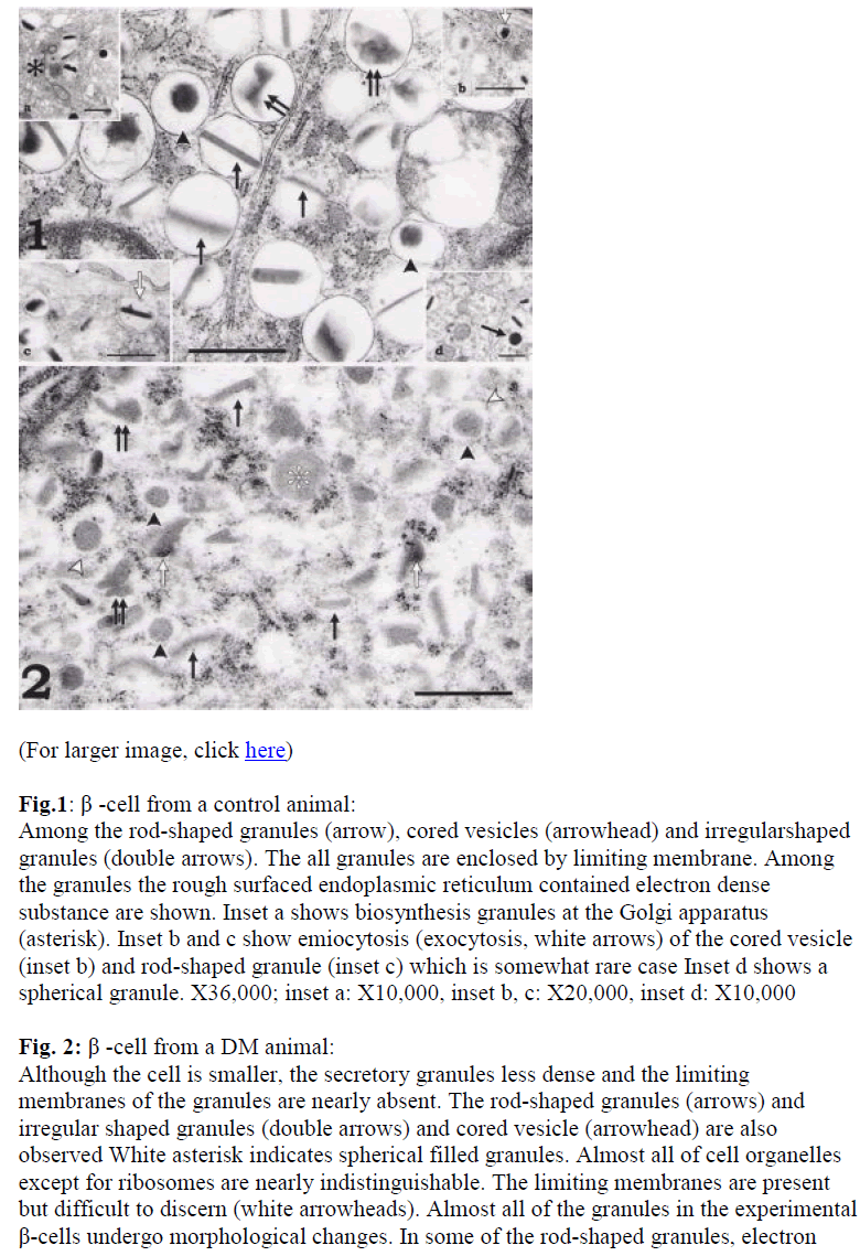
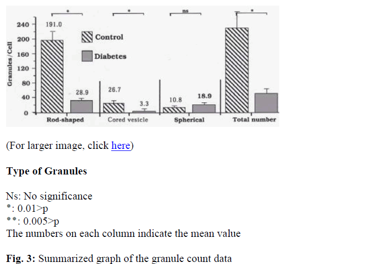
The β-cells of the dog pancreatic islets displayed many rod-shaped secretory granules with a few that were spherical and cored vesicle, so identification of the cell was not difficult (Fig. 1). These granules were mainly located around a well developed Golgi apparatus. Around the Golgi apparatus, the cored vesicle, and spherical granules were also found (Fig.1, inset a). Whereas The granules extrusion of the cord vesicle were often observed (Fig. 1a, inset b), the direct granule extrusion of the rod-shaped granule were observed few (Fig. 1, inset c). No granule extrusion of the spherical granule which was shown in inset d of Fig. 1 was observed. The β-granules in the pancreas of the control dogs showed high electron density. They were, however, less dense and fewer in the DM animals (Fig. 2). A few oval mitochondria were also scattered in the cytoplasm (Figs. 1, 2 and 3).
Fig.1: β -cell from a control animal: Among the rod-shaped granules (arrow), cored vesicles (arrowhead) and irregularshaped granules (double arrows). The all granules are enclosed by limiting membrane. Among the granules the rough surfaced endoplasmic reticulum contained electron dense substance are shown. Inset a shows biosynthesis granules at the Golgi apparatus (asterisk). Inset b and c show emiocytosis (exocytosis, white arrows) of the cored vesicle (inset b) and rod-shaped granule (inset c) which is somewhat rare case Inset d shows a spherical granule. X36,000; inset a: X10,000, inset b, c: X20,000, inset d: X10,000
Fig. 2: β -cell from a DM animal: Although the cell is smaller, the secretory granules less dense and the limiting membranes of the granules are nearly absent. The rod-shaped granules (arrows) and irregular shaped granules (double arrows) and cored vesicle (arrowhead) are also observed White asterisk indicates spherical filled granules. Almost all of cell organelles except for ribosomes are nearly indistinguishable. The limiting membranes are present but difficult to discern (white arrowheads). Almost all of the granules in the experimental β-cells undergo morphological changes. In some of the rod-shaped granules, electron
Type of Granules
Ns: No significance ٭: 0.01>p
٭٭: 0.005>p
The numbers on each column indicate the mean value
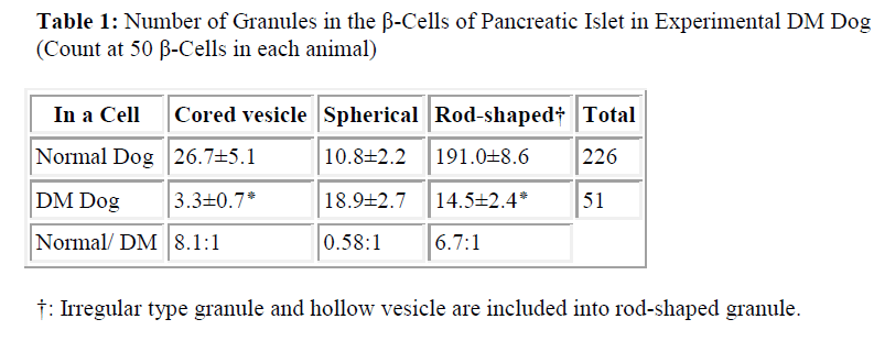
Fig. 3: Summarized graph of the granule count data The number of cored vesicle decrease drastically also about rod-shaped granules between the two animal groups, while the number of spherical granules per cell, however, are slightly increase in the DM dogs. Ns: No significant: *: p<0.01 control and DM dogs was approximately 6.6:1 and of the cored vesicle was 8.1:1. Also in contrast to this ratio of spherical granule was 1:2 (Table 1, Fig. 3). It was very interesting that the ratio of both the rod-shaped and cored vesicle between the control and DM dogs were similar (Fig. 3). It was significant (p<0.01) in the ratio of both the rod-shaped and cored vesicle between the control and DM dogs (Fig. 3).
The granules classified as rod-shaped, spherical, and cored vesicle were counted. Mean values of the number of granules in the β -cells were counted in 50 cells in each animal as summarized on Table 1 and Fig. 3. The total granule number per cell in the control dogs was 225 and in the DM dogs they were 51 (Table 1). Clearly, the granule number in the experimental animals was decreased. Besides the rod-shaped granules in the control dogs was 191 and DM dogs was 28.9, and the cored vesicle in the control dogs was 26.7 and DM dogs was 3.3 /cell (Table 1). In contrast to this the spherical granules in the control and experimental animals were 9.6 and 19.9, respectively (Table 1). The ratio of rod-shaped granules between the dense knots are observed (white arrow). Ribosomes are also seen around the granules. X36,000.
Discussion
There are numerous reports about relationship between a streptozotocin and an oxidant [5,6,7]. They reported that the streptozotocin increased some kind of oxidants. The oxidants were the strong oxidation agents as its name. Asai et al, [2] and the present study showed unclearness of the membrane system of the DM dog β -cell. It was understood that the unclearness of the membrane system was due to oxidation of phospholipids of the membrane system of the cells. Thus the synthetic pathway of the insulin suffered damage. After the treatment of Streptozotocin, some β-cells become necrotic, including those in certain tumors such as inslinoma [8].
Lacy [9], Howell et al [10], Misugi et al [11], Watari [12, 13] and Fujita et al [14] reported on the β-cells of the pancreatic islets of various animals. Watari [13] observed these granules in detail using an ultra-high voltage electron microscope, and classified them into 10 types. He reported that the three-dimensional structure of the granules in the snake contains hexagonal crystalline cores shaped like a rhombic dodecahedron. It is generally known that the snake pancreatic β-cell contains conspicuous rod-shaped granules [13,14] which often show a crystalline structure. In general, crystalline structures have a certain hexagonal [15] or rectangular profile [16] or are rod shaped. In present study the rod-shaped granules containned in the dog β-cell showed a crystalloid structure which was interpreted as representing stability and purity of the molecule. It was believed that the crystals were a storage form of a certain protein [15] such as proinsulin in the dog, snake or human.
Recently Asai et al [2] reported a relationship between the rod-shaped and spherical granules in the dog by using of the ultrastructure of the DM dog pancreas. According to them, the pancreatic β-granules especially rod-shaped granule decreased drastically in DM dog. Comparing with the control and DM dog β-cell, whereas the spherical granule was changed from 16/cell to 12/cell, the rod-shaped granule in a cell was decreased 97/cell to 14.5/cell. The granules extrusion of the spherical granules especially of cored vesicles was frequently observed. No granule extrusion of the rod-shaped granules were observed. They concluded that the spherical granules contained active insulin and the rod-shaped granules contained proinsulin, precursor of active insulin. They concluded that the streptozotocin attacked mainly the pathway of the preproinsulin to proinsulin. The spherical granules were separated into two different categories such as filled spherical and cored vesicle in the present study. The ratio of a change of rod-shaped granule between the control and DM dog was approximately 6.7:1. The ratio of filled spherical granules was approximately 1:2. It was interesting that the cored vesicles in DM dog drastically decreased. The ratio of cored vesicles in control vs. DM dog was approximately 8:1. The ratio of cored vesicle showed good correspondence to the ratio of rod-shaped granules (6.7:1). Although the granules extrusions of cored vesicles were frequently observed, no extrusion figures of filled spherical granules were observed. Few granule extrusion figures were observed as former report [2]. It is commonly accepted that the perfusion of glucose results in degranulation of β-cells [17]. Lacy and his coworkers [9,18,19] and Orci et al [20] reported that the emiocytosis (exocytosis) is the major and probably the only mechanism of insulin secretion. It is reasonable to consider that the rod-shaped granules contained the proinsulin (storage form of the insulin), and cored vesicles contained mainly insulin. According to Asai et al [2], the filled spherical granules also contained insulin molecules. The filled spherical granules probably should be understood as some kind of lysosome. Both lysosome and secretory granules were also made in the Golgi apparatus. It is natural to find the insulin molecules in the lysosome.
In conclusion, from morphological data it is strongly suggested that Streptozotocin mainly acts on the pathway from preproinsulin to proinsulin.
References
- Herman L, Satao T, Fitzgerald PJ. The pancreas. In: Electron Microscopic Anatomy. Kurtz SM (ed.) Chapter 3 Academic press, New York, pp 59-95, 1964.
- Asai Y, Morimoto H, Mabuchi Y, Sakuma E, Shirasawa N, Wada I, Horiuchi O, Sakamoto A, Tanaka C, Herbert D C, Soji T. Influence of experimental diabetes mellitus on secretary granules in β-cells in the dog pancreatic islet. Biomedical Research 2007; 18: in press.
- Elliott WC, Houghton DC, Gilbert DN, Baines-Hunter J, Bennet WM. Experimental gentamicin nephrotoxicity: effect of of Streptozotocin-induced diabetes. J Phamacol Exp Ther 1985; 233: 264-270.
- Soji T, Herbert DC: Intercellular communication between rat anterior pituitary cells. Anat Rec 1989; 224: 523-533.
- Kafl HE, Elkashef HA: Effect of sodium orthovanadate on the urinary bladder rings isolated from normal and hypertensive rats. Pak J Pham Sci 2006, 19: 195-201.
- GoodmanAI, Chader PN, Rezzani R, Schwartman ML, Reqan RF, Rodella L, Turkseven S, Lianos EA, Dennery PA, Abraham NG. Heme oxygenase-2 deficiency contributes to diabetes-mediated increase in superoxide anion renal dysfunction. J Am Soc Nephrol 2006, 17: 1073-1081.
- Alber G, Olukman M, Irer S, Caqlayan O, Duman E, Yilmaz C, Ulker. Effect of vitamin E and C supplementation combined with oral antidiabetic therapy on the dysfunction in the neonatally streptozotocin injected diabetic rat. Diabetes Metab Res Rev, 2006 22: 190-197.
- Chang AY, Diani AR. Chemically and hormonally induced Diabetes Mellitus. In: The Diabetic Pancreas Ed: Volk BW and Arquilla ER, chapter 19, pp 415-422, Prenum Medical Book Company, NY and London 1985
- Lacy PF. Electron microscopic identification of different cell type in the islets of Langerhans of the ginea pig, rat rabbit and dog. Anat Rec 1957; 128: 255-267.
- Howell SL, Kostianovsky M, Lacy PF. A study by electron microscopic radioaurtography. J Cell Biol 1969; 642: 695.
- Misugi K, Howell SL, Greider MH, Lacy PF, Sorenson GD. The pancreatic beta cell. Demonstration with peroxidase labeled antibody technique. Arch Pathol 1970; 89: 97-102.
- Watari N, Hotta Y, Mabuchi Y. Morphological studies on vitamin A-strong cell and its complex with macrophage observed in mouse pancreatic tissue following excess vitamin A administration. Okajima Folia Anat Jpn 1982, 58: 837-858.
- Watari N. Crystalline structures observed in both the exocrine and endocrine pancreatic cells. In: Recent Progress in Electron Microscope of Cell and Tissue. Yamada E, Mizuhira V, Kurosumi K, Nagano T (ed), Igaku Shoin Ltd. Tokyo, pp 109; 1976.
- Fujita T, Takamatsu K, Tokunaga J. Pancreatic Islets. In: SEM Atlas of Cells and Tissues. Igaku Shoin, Tokyo, pp 306-310, 1981.
- Fawcett DW. Cytoplasmic inclusions: In: The Cell, second edition. W.B. Saunders Company, Philadelphia pp 668-685, 1981.
- Soji T, Fujita T, Yoshimura F. Crystals in Hyperfunctioning rat parathyroid cells. Endocrinol Japon 1974; 21: 551-553.
- Brown EM Jr, Dohan FC, Freedman LR, De Moor P, Jukens FD. The effect of prolonged infusion of the dog’s pancreas with glucose. Endocrinology 1952; 50: 644-656.
- Lacy PF. Beta cell secretion—from the standpoint of pathobiologist. Diabetes 1970; 19: 895-905.
- Lacy PF. Endocrine secretory mechanisms. A review. Am J Pathol 1975; 79: 170-187.
- Orci L, Amherdt M, Malaisse-Lagae F, Rouiller C. Renold AE. Insulin release by emiocytosis: demonstration with freeze-etching technique. Science 1973; 179: 82-84.


