ISSN: 0970-938X (Print) | 0976-1683 (Electronic)
Biomedical Research
An International Journal of Medical Sciences
- Biomedical Research (2016) Volume 27, Issue 2
Influence of recombinant human Endostatin on the expression of ERCC1 in human lung adenocarcinoma A549 cells.
| Yu Xuan Che, Yan Yan, Xiu Hua Sun* Department of Medical Oncology, The Second Affiliated Hospital of Dalian Medical University, Dalian 116027, Liaoning Province, PR. China |
| Corresponding Author: Xiu Hua Sun, Department of Medical Oncology, The Second Affiliated Hospital of Dalian Medical University, Shahekou District, Liaoning Province, PR. China |
| Accepted: January 20, 2016 |
Objective: To investigate the effect of Endostar and/or DDP on the expression of ERCC1 gene and to provide help to the secondary treatment of lung cancer.
Methods: Human lung adenocarcinoma A549 cells were treated by DDP and rh-Endostatin. MTT method was used for detecting growth inhibition of A549 cells by Rh-Endostatin, DDP and combination of both drugs to determine the applied concentration.
Results: 1. By MTT method,Rh-Endostatin had no cytotoxic effect on A549 cells at the concentration of 300 μg/ml. IC30 of DDP to A549 cells was 0.34 μg/ml, and IC30 of DDP in combination with Rh-Endostatin to A549 cells was 0.13 μg/ml. 2. By real time PCR, mRNA relative expression of ERCC1 genes in each group were as follows: (i)1.00; (ii)0.88; (iii) 2.00; (iv)1.90; (v)0.77.The resulting: (1) Expression in all treatment groups was higher than that in control group. (2) Expression in combination treatment groups was higher than those in single-drug treatment group.
Conclusions: 1. Cytotoxic effect of DDP to lung adenocarcinoma A549 cells is enhanced by Rh-Endostatin. 2. Expression of ERCC1 mRNA in A549 cells is enhanced in combination treatment by DDP and Rh-Endostatin.
Keywords |
||||||||||||||||||||||
| Lung cancer; Recombinant human endostatin; Cisplatin; Excision repair cross complement-1; A549 cells. | ||||||||||||||||||||||
Introduction |
||||||||||||||||||||||
| Lung cancer is a common lethal malignancy around the world and the incidence of lung cancer has increased significantly year by year. The treatment for lung cancer is based on the comprehensive therapies and chemotherapy on the basis with cisplatin (DDP) plays an important role. However, the occurrence of multidrug resistance (MDR) can result in the failure of chemotherapy through the nucleotide excision repair (NER) pathway. Excision repair cross complement-1 (ERCC-1) is the rate-limiting enzyme of NER pathway and the overexpression of ERCC1 could rapidly repair the damaged DNA stuck at the G2/M phase. The ERCC1 expression correlates with the mechanism of DDP resistance for the treatment of lung cancer [1-3]. Since 1971, Folkman et al. proposed the hypothesis that tumor cells relied on the formation of tumor blood vessels and anti-tumor vessels treatment has become the research hotspot [4]. Recombinant human Endostatin (rh-Endostatin) can not only block the vascular endothelial growth factor (VEGF) pathway, but also inhibit the formation of tumor vessels through the nucleolin [5-6]. Our experiment suggested that rh-Endostatin can enhance the inhibition of DDP on the A549 cells and the increase expression of ERCC1, which may provide a new approach for the second-line of lung cancer therapy. | ||||||||||||||||||||||
Materials and Methods |
||||||||||||||||||||||
Materials |
||||||||||||||||||||||
| Human lung adenocarcinoma A549 cells were purchased from Science cell Co. (USA). The PRMI 1640 culture was supplied by Hyclone Co. (USA). The MTT was obtained from Ameresco Co. (USA). And DMSO and trypsin were purchased from Sigma Co. (USA). The rh-Endostatin was obtained by Xiansheng pharmacy Co. (Jiangsu, China). Cisplatin was purchased from Haosen pharmacy Co. (Jiangsu, China). Fetal bovine serum (FBS) was supplied by TBD biological Co. (Tianjin, China). AnnexinV-FITC apoptosis kit was purchased from Biouniquer Co. (USA). DMSO and trypsin were purchased from Sigma Co. (USA).DL 2000 DNA Marker and Diluent were prepared for Real-time PCR. | ||||||||||||||||||||||
Tumor cell preparation |
||||||||||||||||||||||
| The human lung adenocarcinoma A549 cells were intensively cultured in the PRMI 1640. After the cryopreservation and resuscitation, the cancer cells were then used for culturing in PRMI 1640 until they reached 80% confluence. Finally, the cancer cells were washed in PBS solution. And when the shape of cells turned round and there was gap between cells, the reaction was terminated by adding complete medium. Single cell suspension was prepared and then passaged as a ratio of 1:2 or 1:3. | ||||||||||||||||||||||
Detection of cisplatin, rh-Endostatin and the combination of cisplatin and rh-Endostatin on the inhibition of A549 cells proliferation |
||||||||||||||||||||||
| The cancer cells were plated in 96-chamber culture plates at 20,000 cells per well for 24 h, allowed to adhere and incubate with the different concentration of cisplatin all at once or in three days, rh-Endostatin and the combination of cisplatin and rh-Endostatin for 72 h respectively. And then the MTT liquid was added to each plate and culture for 4 h. Absorbance (A value) at 570 nm was measured with Model 550 Type microplate reader after dissolving with DMSO. And the inhibition rate was calculated to determine the inhibition effects of antitumor drugs on tumor cells. Absorbance was detected repeatedly 3 times on different days and expressed as mean±standard deviation. | ||||||||||||||||||||||
Detection of ERCC1mRNA on the A549 cells through real-time PCR |
||||||||||||||||||||||
| The cancer cells RNA were purified and DNA were removed and conducted reverse transcription.And then we selected the β-actin as housekeeping gene,which the forwardprimer was 5’- TGGCACCCAGCACAATGAA-3’ and the reverseprimer was 5’-CTAAGTGATAGTCCGCCTAGAAGCA-3’ with the amplification length of 186bp. The forwardprimer of ERCC1 was 5’-GGAGACCTACAAGGCCTATGAGCA-3’ and the reverseprimer of ERCC1 was 5’- ACTTCACGGTGGTCAGACATTCAG-3’ with the amplification length of 104 bp. And then conducted the polymerase chain reaction. | ||||||||||||||||||||||
Statistical analysis |
||||||||||||||||||||||
| The statistical analysis was performed with SPSS 16.0 software and statistical differences were determined by oneway ANOVA. The data were expressed as the mean ± standard deviation and all experiments were performed in twice. The value of P<0.05 was considered to indicate a statistically significant difference. | ||||||||||||||||||||||
Results |
||||||||||||||||||||||
| • The inhibition of different drugs on the growth of human lung adenocarcinoma A549 cells by MTT approach. | ||||||||||||||||||||||
| The results showed that the rh-Endostatin can inhibit the growth of A549 cells and the inhibition rate improved with the increase of the concentration. And the rh-Endostatin with 300 μg/ml had no cytotoxicity on the A549 cells,so we chose the rh-Endostatin with 300 μg/ml as the experiment concentration (Table 1). | ||||||||||||||||||||||
| • The analysis of relative quantity of ERCC1 mRNA by real time PCR | ||||||||||||||||||||||
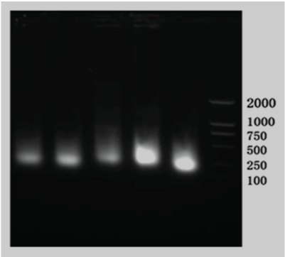
| • The purity and quality of total RNA (Figure 1). | ||||||||||||||||||||||
| • The results of real time PCR | ||||||||||||||||||||||
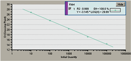
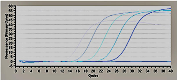
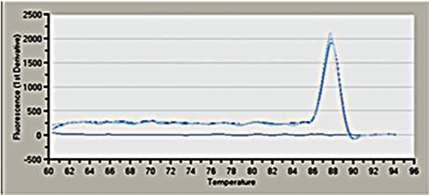
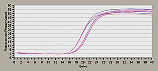
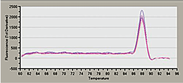
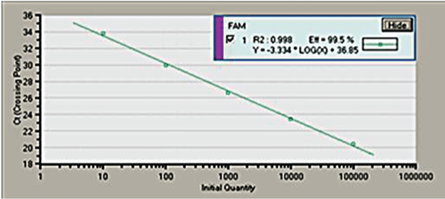
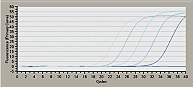
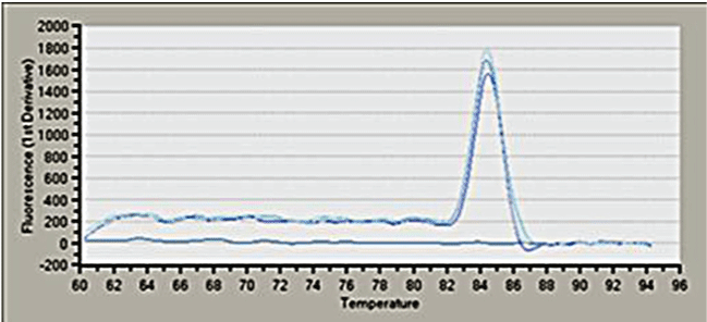
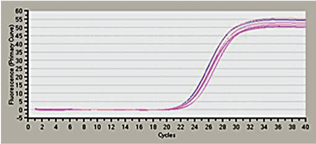
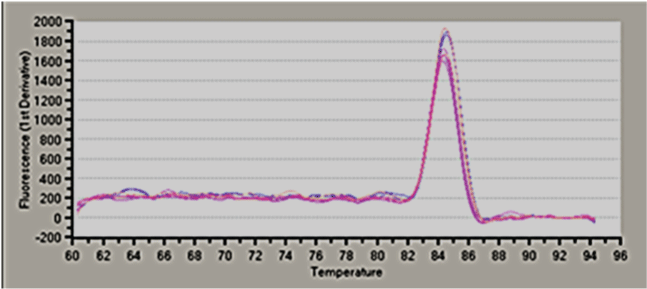
| • The standard curve of housekeeping gene β-actin (Figures 2-11). | ||||||||||||||||||||||
| Chemotherapy on the basis of DDP is the first-line regimen for the treatment of lung cancer [7]. DDP can promote the death of cancer cells through the combination with DNA which inhibit the DNA reproduction and DNA transcription [8]. The drug resistance of DDP plays an important role in the chemotherapy failure for non-small cell lung cancer (NSCLC). If damaged DNA can repair in time, the cancer cells were still resistant to DDP which results in the chemotherapy failure [9,10] (Table 2). | ||||||||||||||||||||||
| The NER pathway is the main way for the DNA repair in mammalian cells which is also the only mechanism for eliminating the helixdistorting DNA caused by DDP [11]. NER pathway is a complex progress including the recognition, incision, resection, synthesis and connection which takes part in the damaged DNA repair caused by the ultraviolet rays or chemicals in order to maintain the stability of genome [12]. NER proteins include two groups. ERCC1 is one of the nucleotide excision repair enzymes family which takes an important position in the DNA recognition and inter-chain incision in the NER pathway and the activity of ERCC1 can reflect the level of the NER repair activity. In some tumor cells,the low expression of ERCC1 mRNA indicates that the nucleotide excision repair decreased which results in the occurence of cancer [13]. The over-expression of ERCC1 could rapidly repair the damaged DNA stuck at the G2/M phase.,especially increase the elimination of DNA compound induced by DDP [14]. Zhou et al. demonstrated that the inhibition of ERCC1 expression can reduce the repair of DDPDNA complex which may decrease the resistance of tumor cells [15]. Ryu et al. suggested that patients with 118 codon C/C genotype on the ERCC1 gene were more likely to benefit from the chemotherapy with DDP and less resistance than patients with other genotype [16]. | ||||||||||||||||||||||
| Our experiment discovered that the expression of ERCC1 mRNA increased slightly on the A549 cells by once exposure chemotherapy with DDP and continous exposure chemotherapy with DDP than the control group, which may indicate that the possibility of resistance with the long-term use of DDP. What is more, there is no significant differences between the once exposure chemotherapy with DDP and continuous exposure chemotherapy on the ERCC1 expression (P>0.05), which indicates that the occurance of DDP resistance is not relied on the drug administration. | ||||||||||||||||||||||
| Since 1971, Folkman et al. proposed the hypothesis that tumor cells relied on the formation of tumor blood vessels. And some researchers proposed that the formation of tumor blood vessels takes an important in the invasion and metastasis of tumor cell. Thus anti-tumor vessels treatment has become the research hotspot. | ||||||||||||||||||||||
| Rh-Endostatin is an endogenous angiogenesis inhibitors developed by China which can target more than 12% angiogenesis regulatory gene of human genome. Rh-Endostatin can inhibit the angiogenesis of tumor, and block nutrient supply to tumor cells. So rh-Endostatin can achieve the antitumor effects by inhibiting the migration and proliferation of endothelial cells and inducing apoptosis of vascular endothelial cells. | ||||||||||||||||||||||
| In our experiment, we discovered that the amount of A549 cells death in the combination of DDP and rh-Endostatin with non-cytotoxicity group is much more than the DDP group, which may indicate that rh-Endostatin can increase the chemotherapy sensitivity. Through the MTT approach, we also discovered that the IC30 of A549 cells decreased significantly in the combination of DDP and rh-Endostain than the single DDP group, which also indicate that the combination can increase the effect of chemotherapy. | ||||||||||||||||||||||
| We chose the combination of DDP and rh-Endostatin on the A549 cells in once exposure and continous exposure and the results showed that the expression of ERCC1 mRNA increased significantly in the combination group than the control group. According to the results, we indicated that the combination of DDP and rh-Endostatin for long-term treatment may result in the resistance of DDP and the more possibility of resistance in the combination of DDP and rh-Endostatin than the DDP single (P<0.05). We think that most of the cancer cells sensitive to the chemotherapy has been eliminated with the combination, so the rest of cancer cell may become the cells with resistance, which result in the expression of ERCC1 increased. However, we have not observed for long-term administration, but the short-time administration on the expression of ERCC1. So the mechanism of the phenomenon is still to be investigated. | ||||||||||||||||||||||
Conclusions |
||||||||||||||||||||||
| • Cytotoxic effect of DDP to lung adenocarcinoma A549 cells is enhanced by Rh-Endostatin. | ||||||||||||||||||||||
| • Expression of ERCC1 mRNA in A549 cells is enhanced in combination treatment by DDP and Rh-Endostatin. | ||||||||||||||||||||||
Acknowledgement |
||||||||||||||||||||||
| This study is supported by Natural Science Foundation of Liaoning Province, No.201202043. | ||||||||||||||||||||||
Tables at a glance |
||||||||||||||||||||||
|
||||||||||||||||||||||
Figures at a glance |
||||||||||||||||||||||
|
||||||||||||||||||||||
References |
||||||||||||||||||||||
|
