ISSN: 0970-938X (Print) | 0976-1683 (Electronic)
Biomedical Research
An International Journal of Medical Sciences
- Biomedical Research (2016) Volume 27, Issue 2
Intrarenal angiotensin II plays an important role in renal fibrosis in primary IgA nephropathy.
| Han Wen-lun*, Zhu Yun-yun, Zhong Yu, Ding Hui-deng Department of Nephrology, Tongde Hospital of Zhejiang Province, PR. China |
| *Corresponding Author: Han Wen-lun,Department of Nephrology Tongde Hospital of Zhejiang Province PR. China |
| Accepted: January 20, 2016 |
Objective: To investigate the relationship of intrarenal angiotensin II (Ang II) expression with clinicpathological injury index and intrarenal expression and regulation of RAS (renin-angiotensin system) components in IgAN (immunoglobulin A nephropathy) patients.
Material and methods: The expression of intrarenal renin, angiotensinogen (AGT), Ang II, Ang IIreceptor was examined by immunohistochemistry staining (IHCS). Clinic-pathological injury index included blood pressure, estimated glomerular filtration rate (eGFR), proteinuria and Katafuchi score.
Results: Positive IHCS area of intrarenal Ang II was 32.73 ± 14.74%. There was a negative correlation between intrarenal Ang II and eGFR (r=-0.61, P<0.01), index of glomerular hypercellularity (ρ=-0.35, P<0.05).Intrarenal Ang II correlated positively with pathological chronicity index (ρ=0.39, P<0.05), index of interstitial cell infiltration (ρ=0.52, P<0.01), index of tubular atrophy (ρ=0.51, P<0.01), index of vessel wall thickening (ρ=0.36, P<0.05) and index of arteriolar hyalinosis (ρ=0.36, P<0.05). Intrarenal Ang II positively correlated with intrarenal renin and AGT (r=0.43, P<0.01 and r=0.3, P<0.05, respectively). No other correlation was evidenced between intrarenal expression of explored RAS components.
Conclusions: Intrarenal Ang II plays an important role in renal fibrosis in IgAN, particularly at the tubulointerstitial level. Moreover, there is a tight regulation of the intrarenal RAS components in IgAN.
Keywords |
||||||
| IgA nephropathy, Renin-angiotensin system, Estimated glomerular filtration rate, Katafuchi score, Chronicity. | ||||||
Introduction |
||||||
| A number of studies have shown that activation of intrarenal renin-angiotensin system (RAS) plays an important role in development and progression of chronic kidney disease (CKD). Surprisingly, little information is available about the RAS expression and regulation in the human kidney and particularly in kidney diseases and data on the intrarenal RAS expression and regulation were mostly obtained in animals [1,2]. These data in humans and in diseased kidneys would be worthwhile evaluating because changes in general RAS do not closely reflect local expression and regulation of intrarenal RAS [3]. On the contrary, the simultaneous assessment of the expression and regulation of all components of intrarenal RAS is necessary for evaluation of the net effect of RAS on the kidney. Indeed, the effect of RAS on the kidney cannot be accurately assessed by the measurement of one component alone. For instance, the local availability and functional consequence of angiotensin II (Ang II), the bioactive substance of the system, may depend on the Angiotensin converting enzyme (ACE) concentration or on the Ang II receptor density [4]. The present study was carried out to obtain deeper insight on the RAS regulation in the diseased kidneys in view of the pathogenic role of this system, particularly in the progression to renal failure [5,6]. As a paradigmatic renal disorder, we investigated immunoglobulin. A nephropathy (IgAN), the most common type of primary glomerulonephritis worldwide and a major cause of End stage renal failure (ESRF). Expression of RAS components were simultaneously investigated by Immunohistochemistry staining (IHCS). | ||||||
Material and Methods |
||||||
Patients and samples |
||||||
| 72 Chinese primary IgAN patients who had undergone renal biopsy in Zhongshan Hospital between January 2009 and June 2009, had not received glucocorticoid, immunosuppressants, angiotensin converting enzyme inhibitor or angiotensin IIreceptor blocker, and gave informed consent were included in the study. The study protocol was approved by the ethics committee of Zhangshan Hospital, Fudan University. The patients included 20 men and 52 women with a mean age of 35.67 yr (range 23 to 60 yr). The estimated glomerular filtration rate (eGFR) ranged from 6.62 to 92.49 ml/min/1.73 m2 (mean 55.92 ml/min/1.73 m2). The eGFR was calculated using the Modification of Diet in Renal Disease Formula (eGFR=175 × standardized serum creatinine-1.154 × age-0.203 × 0.741 [if Asian] × 0.742 [if female]) [7], which was found to correlate well with GFR corrected for body surface area in adults. | ||||||
| Renal biopsies samples were obtained under ultrasound guidance with a 16-gauge needle. Only those biopsies disclosing the typical immunofluorescence for IgAN and providing a sufficient sample for performing both the standard pathologic examination and molecular biology analysis were included for this study. | ||||||
Renal immunohistochemistry staining |
||||||
| We performed IHCS for renin, Angiotensiongen (AGT), Ang II, Angiotensin II type 1 receptor (AT1R), Angiotensin II type 2 receptor (AT2R) in consecutive kidney section. Microwave irradiation was performed to enhance antigen retrieval. The primary antibodies were sheep anti-human renin (R&D systems, US), rabbit anti-human angiotensinogen (Sigma- Aldrich, St. Louis, MO, USA), rabbit anti-human angiotensin II (Phoenix pharmaceutics, Burlingame, CA, USA), mouse anti-human AT1R (Abcam Ltd, Cambridge, UK), rabbit antihuman AT2R (Abcam Ltd, Cambridge, UK). The secondary antibodies were donkey anti-rabbit IgG (DakoCytomation Ltd, Cambridge, UK), donkey anti-mouse IgG (DakoCytomation Ltd, Cambridge, UK) and rabbit anti-sheep IgG (KPL, Gaithersburg, Maryland, USA). A schematic representation is shown in Table 1. Sections that were incubated with phosphate buffered saline, as appropriate, instead of the primary antibodies served as negative controls. All sections were stained under identical conditions together with control incubation. The immunoreactivities were scored twice in a blind manner by Leica QWin V3 imaging analysis software and the interassay variations were not significant. | ||||||
Histopathologic evaluation |
||||||
| One of the investigators reviewed all the histologic specimens and for each biopsy three serial sections stained with hematoxylin-eosin, periodic-acid Schiff (PAS), and Masson’s trichrome were evaluated. The overall severity of renal damage was graded according to the Katafuchi score [8]. In each biopsy eight features were assessed: glomerular hypercellularity (1 ~ 4), glomerualr segmental lesions (0 ~ 4), glomerualr sclerosis (0 ~ 4), interstitial fibrosis (0 ~ 3), interstitial cell infiltration (0 ~ 3), tubular atrophy (0 ~ 3), vessel wall thickening (0 ~ 3) and arteriolar hyalinosis (0 ~ 3). Pathological activity index was defined as the summary of glomerular hypercellularity, glomerualr segmental lesions, interstitial cell infiltration and arteriolar hyalinosis. Chronicity index was defined as the summary of glomerualr sclerosis, interstitial fibrosis, tubular atrophy and vessel wall thickening. The histopathologic score was done in a blind manner by two independent pathologists and the interassay variations were not significant. | ||||||
Statistical analysis |
||||||
| Statistics were carried out by linear regression analysis of different intrarenal RAS components expression and clinicpathological injury index. We conducted Pearson and Spearman single regression analysis for parametric and nonparametric data, respectively, among all parameters studied. Statistical significance was set at P<0.05. Statistical analysis was performed with SPSS software, version 17.0. | ||||||
Results |
||||||
Relationship between intrarenal angiotensin II and clinic-pathological injury index in IgA nephropathy |
||||||
| Positive IHCS area of intrarenal Ang II was 32.73 ± 14.74% (6-70%). A negative correlation emerged between intrarenal Ang II and eGFR(r=-0.61, P<0.01), index of glomerular hypercellularity (ρ=-0.35, P<0.05). On the contrary, intrarenal Ang II and pathological chronicity index (ρ=0.39, P<0.05), index of interstitial cell infiltration (ρ=0.52, P<0.01), index of tubular atrophy (ρ=0.51, P<0.01), index of vessel wall thickening (ρ=0.36, P<0.05) and index of arteriolar hyalinosis (ρ=0.36, P<0.05) were linked through a positive relationship. A schematic representation is shown in Table 2. | ||||||
Intrarenal expression and regulation of RAS components in IgA nephropathy |
||||||
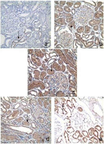
| Positive IHCS area of intrarenal renin, AGT, Ang II, AT1R and AT2R were 26.86 ± 13.66% (7-55%), 38.34 ± 9.71% (12-57%), 32.73 ± 14.74% (6-70%), 36.67 ± 16.78% (6-81%) and 28.43 ± 14.66% (4-78%), respectively. Renin staining is detected mainly in juxtaglomerular apparatus and tubular cells (Figure 1a). AGT staining is detected mainly in tubular cells and some glomerular cells (Figure 1b). Ang II staining is detected mainly in tubular cells and some glomerular cells (Figure 1c). AT1R staining is detected mainly in tubular cells and some glomerular cells (Figure 1d). AT2R staining is detected mainly in tubular cells (Figure 1e). Intrarenal Ang II positively correlated with intrarenal renin and AGT (r=0.43, P<0.01 and r=0.34, P<0.05, respectively). No other correlation was evidenced between explored RAS components. A schematic representation is shown in Table 3. | ||||||
Discussion |
||||||
| Recently studies have demonstrated that immunoreactivity of AGT in tubules was significantly increased in IgAN patients compared to the control group [9]. Immunoreactivity of AGT was significantly correlated positively with urinary occult blood, urinary protein-to-creatinine ratio, urinary protein excretion and serum creatinine [9]. Immunoreactivity of angiotensinogen was also significantly correlated negatively with creatinine clearance [9]. It was reported that RAS genes were overexpressed in IgAN and a tight regulation of the intrarenal RAS existed by glomerular and tubulointerstitial compartments microdissection and reverse transcriptionpolymerase chain reaction (RT-PCR) [4]. There was no difference between glomerular and tubulointerstitial RAS gene expression levels. On the contrary, the overactivation of fibrogenic cascade genes, such as transforming growth factor-1 (TGF-ß1), collagen IV (Coll IV), a-smooth muscle actin(a- SMA), in the tubulointerstitium was observed. This fibrogenic cascade seems to be triggered by RAS as indicated by statistically significant correlations between the expression of their respective genes [4]. | ||||||
| A direct relationship between the expression of AT receptor genes and the putative Ang II activity was found in the tubulointerstitium, while in the glomeruli the association was negative. In the interstitium, interstitial infiltrates was related to the gene expression of Agtg, AT1 receptor, Coll IV, and TGF-beta1positively [4]. Actually Ang IIincreases monocyte adhesion to the endothelium and is chemotactic for neutrophil leukocyte [10] and monocyte/macrophage [11,12] and a role of the RAS in renal inflammation has been proposed. The in situ hybridization study researched by Lai et al. [13] which did not report RAS gene expression in inflammatory cells in IgAN supported the idea that interstitial infiltrates could be a phenomenon secondary to RAS activation. Our results demonstrated that intrarenal Ang IIwas positively correlated with both renin and AGT by immunohistochemistry staining. These data suggests that intrarenal RAS in human IgAN is strictly regulated as demonstrated by the correlation between different components of the RAS cascade. | ||||||
| Moreover, the expression of intrarenal Ang IIis strictly correlated with some clinic-pathological injury index in IgAN. It is conceivable that increased intrarenal Ang IIIHCS area with low eGFR and high pathological chronicity index is mediated by enhanced intrarenal Ang IIactivity and results in progressive renal fibrosis and progressive deterioration of renal function. At the same time, intrarenal Ang IIis positively correlated with index of interstitial cell infiltration, tubular atrophy, vessel wall thickening, arteriolar hyalinosis and inversely correlated with the index of glomerular hypercellularity. We speculate that such a correlation may indicate intrarenal Ang IIplay an important role in interstitial and vascular lesion, especially chronic interstitial and vascular lesion. Dorella et al. [4] also observed that fibrogenic cascade genes (TGF-ß1, Coll IV, a-SMA) in the tubulointerstitium significantly correlated with RAS genes in IgAN. Indeed, one of the most robust predictors of poor prognosis in human nephropathies, including IgAN, is interstitial fibrosis and inflammation rather than glomerular damage [14]. Overexpression of intrarenal Ang IImay predict poor prognosis in IgA nephropathy. | ||||||
| In conclusion, this study demonstrates that intrarenal Ang IIseems to play an important role on renal fibrosis in IgAN, particularly at the tubulointerstitial level, which has a significant impact on the prognosis in human nephropathies. Moreover, a tight regulation of the intrarenal RAS components exists in IgAN. These findings confirm the rationale for the early treatment with angiotensin converting enzyme inhibitor or angiotensin IIreceptor blocker in IgAN even in the absence of hypertension and/or severe proteinuria. However, there were still some limitations in this research. First of all, whether the duration of the disease could affect the results was unknown as few participants were included in this research and we could not perform the hierarchical analysis. Furthermore, the research was conducted in china. Whether the conclusion was suitable for person from the world was unknown. Meanwhile, this research was conducted in a single center, multicenter clinical study was needed for further exploration. At last, the study drawn only a simple correlation conlusion but not a causal relationship. Whether the immnological pathogenesis of IgAN results in or from the intrarenal angiotensinIIactivation still needs further cohort or in vitro study. | ||||||
Acknowledgements |
||||||
| This work was funded by the Science and Technology Commission of Shanghai (08DZ1900602) and Key Subject Construction Project (Phase 3), the National ‘211 Project’, Ministry of Education, China. | ||||||
Tables at a glance |
||||||
|
||||||
Figures at a glance |
||||||
|
||||||
References |
||||||
|
