ISSN: 0970-938X (Print) | 0976-1683 (Electronic)
Biomedical Research
An International Journal of Medical Sciences
- Biomedical Research (2013) Volume 24, Issue 3
Investigating relative strengths and positions of electrical activity in the left and right hemispheres of the human brain using Electroencephalography.
Symbiosis Institute of Technology, Symbiosis International University, Pune, Maharashtra, India
- Corresponding Author:
- Meena Laad
Department of Applied Science
Symbiosis Institute of Technology
Symbiosis International University Symbiosis Knowledge Village
Gram:Lavale Pune, India
Accepted date: April 19, 2016
Citation: Laad M. Investigating relative strengths and positions of electrical activity in the left and right hemispheres of the human brain using Electroencephalography. Biomed Res-India; 24 (3):359-364.
The present study determines the relative strengths and positions of electrical activity in different regions of the brain. It aims to investigate the differential involvement of the left and right hemispheres of the brain when a visual stimulant is administered. This paper examines the frequency - intensity spectrum of recorded EEG signals at the electrodes at F7, F8, P3, P4, T3, T4, O1, O2, C3, C4, T5 and T6 situated in two hemispheres at a specific instant and inferences were drawn regarding hemispheric differences in EEG activities in four frequency bands 0-4 Hz, 4-8 Hz, 8-12 Hz and 12-25 Hz for all the four subjects. The present work will be useful for future research to study the importance of the functional asymmetry of the brain for the organization of emotions though the relative contribution of the left and right hemispheres. The findings of this study can also be used to develop brain-computer interface for various applications.
Keywords
Brain-computer interface, electroencephalography, frequency, intensity.
Introduction
Electroencephalography is a medical imaging technique that records scalp electrical activity generated by different regions in the brain. Brain electrical activity is generated by electric volume conductor i.e. the cortex, skull, meninges, and scalp [1]. With the latest development in the field of signal analysis EEG has emerged as a brain mapping and brain imaging technique with great potential to providing important spatio-temporal information regarding brain. EEG also has the benifit of being compatible with other brain imaging techniques. Brain imaging techniques allow researchers and scientists to view activity at various regions of human brain, without invasive neurosurgery. There are a number of established, safe imaging techniques in use today throughout the world. Each of these brain imaging techniques has their own unique strengths and weaknesses, and there may be potential benefits and difficulties in combining these techniques to achieve a complete analysis of brain functioning. The most accepted brain imaging techniques are Magnetic Resonance Imaging (MRI), Near InfraRed Spectroscopy (NIRS), Positron emission tomography (PET), SPECTSingle Photon Emission Computed Tomography, EEGElectroencephalography. MRI makes use of strong magnetic fields and electromagnetic pulses to excite protons which then generate a photon before decaying to their normal state. These photons are then measured by the MRI and a map of a living tissue is produced. Functional MRI depends on the magnetic properties of blood and enable researchers to see images of blood flow in the brain as it is occurring. In NIRS a near Infra red light source and an NIR light detector are attached to the scalp and the transmission and absorption rate of the NIR light in human tissues is obtained, which contains information of the changes in haemoglobin concentration. The oxygen demand increases when a particular area of the brain is active and thus haemoglobin concentration also increases. The NIRS are portable and easy to use in comparison with MRI. In Positron Emission Tomography (PET) the subject under study is injected with a radioactive marker that contains isotopes that decays into lower energy particles, and create positrons which collide with electrons and then transform into photons that can be detected by the PET. This brain imaging technique is invasive it is expensive and is not portable. But the quality of PET images is very high and is widely used for Brain Tumour detection, and various other clinical applications.
SPECT-Single Photon Emission Computed Tomography uses radioactive tracers and a scanner to record data that a computer uses to construct two- or three-dimensional images of active brain regions. EEG-Electroencephalography uses electrodes placed on the scalp to detect and measure patterns of electrical activity emanating from the brain. EEG can determine the relative strengths and positions of electrical activity in different brain regions. The greatest advantage of EEG is speed-it can record complex patterns of neural activity occurring within fractions of a second after a stimulus has been administered [2]. EEG data contains useful information about physiological, psychological and pathological processes as different regions in the brain reflect different EEG data. The information derived by analyzing and processing EEG data can be used for the diagnosis of brain disorders, psychological behavior and various brain related research applications. Electroencephalography is a noninvasive and inexpensive technique with very high sensitivity to receive information about the internal structure and functioning of the brain. It’s being used for various applications in Cognitive Development and Psychological research studies. The general cognitive processes involve perception, memory, attention, language, and emotion in normal adults and children. It provides a very high time resolution in the millisecond range [3]. EEG data can be effectively used for Brain Computer Interface applications for establishing a new channel for communication between human brain and computers. The greatest advantage of EEG is its high speed. Complex patterns of neural activity occurring within fractions of a second can be recorded with EEG after administering a stimulus [4]. The most useful application of EEG recording is the ERP (event related potential) technique. It is well known fact that the EEG signals are the measure of the alertness of brain which changes according to task performed by a person. The brain waves can be classified into few different frequency bands named as delta, theta, alpha, beta and gamma. The accurate classification of electrical activity for a particular state of human brain helps in neurological diagnosis and also for establishing standards for developing proper techniques and instruments. This classification can be utilized in developing brain computer interface applications.
The human brain is divided into two equal halves, left hemisphere and right hemisphere. According to functions performed, each half is divided into four main lobes as mentioned below:
1. Frontal lobe: The frontal lobes play important role in impulse control, judgment, language production, working memory, motor function, sexual behavior, socialization and spontaneity. The frontal lobe assists in planning, coordinating, controlling and executing behavior.
2. Occipital Lobe: The visual cortex is located in the occipital lobe. The occipital lobe is responsible for one’s ability to see and visual interpretation.
3. Parietal Lobe: The parietal lobe helps in integrating sensory information from different parts of the body, knowledge about numbers and the relation between them, and in the manipulation of the objects.
4. Temporal Lobe: The temporal lobes deal primarily with memory functions. The dominant temporal lobe is specialized for verbal (word-based) memory and the names of objects. We depend upon our non-dominant temporal lobe for our memory of visual (non-verbal) material, such as faces, scenes.
EEG electrode Positioning
In EEG recording, each scalp electrode is placed near certain brain centres because different areas are responsible for different functions in the brain e.g. F7 is located near centres for rational activities, Fz near intentional and motivational centres, F8 close to sources of emotional impulses. Cortex around C3, C4, and Cz locations deals with sensory and motor functions. Locations near P3, P4, and Pz contribute to activity of perception and differentiation. Near T3 and T4 emotional processors are located, while at T5, T6 certain memory functions stand. Primary visual areas can be found below points O1 and O2. However the scalp electrodes may not reflect the particular areas of cortex, as the exact location of the active sources is yet to be found out due to limitations imposed by the nonhomogeneous properties of the skull, different orientation of the cortex sources, coherences between the sources, etc [5].
While positioning the electrodes on the scalp, there needs to be a reference electrode with respect to which the voltage of EEG signals at other electrodes will be measured. Several different recording reference electrode placements are mentioned in the literature. Physical references can be chosen as vertex (Cz), linked-ears, linkedmastoids, ipsilateral-ear, contralateral-ear, C7 reference, bipolar references, and tip of the nose. Electrode positions are named according to the brain region below the area of the scalp: frontal, central, parietal, temporal, and occipital. In the bipolar method, the EEG is measured from a pair of scalp electrodes. The pair of electrodes measures the electrical potential difference between their two positions above the brain with respect to a reference electrode. There are reference-free techniques represented by common average reference, weighted average reference, and source derivation. To prevent signal distortions impedances at each electrode contact with the scalp is kept below 5 KΩ.
Materials and Method
For our current study, EEG data were recorded from two male and two female subjects in the age group of four to fourteen years. None of them had any medical history or had taken any medication that could have affected the EEG signal. The subjects were made to sit comfortably on a chair and EEG electrodes were fixed on the scalp as per International 10-20 electrode placement system. The visual stimulus in the form of a pleasant picture was shown to the subjects and EEG data was recorded. The frequency analysis for all the four subjects was done at 60th second from the start. The signal intensity at electrodes F7, F8, P3, P4, T3, T4, O1, O2, C3, C4, T5 and T6 was calculated and tabulated for four frequency bands 0 - 4 Hz, 4 - 8 Hz, 8 - 12 Hz and 12 - 25 Hz. The variation in the intensity at each electrode placed in left and right hemisphere were plotted for each frequency band for subjects A, B, C and D separately to observe the response of each electrode for the same stimulus and to study the behavior of different regions in the brain in the left and right hemisphere. This study also aims to understand the response of different regions of brain for subjects of different background and age for the same stimulus.
Experimental Observations
The recorded waveforms reflected the cortical electrical activity. EEG activity is measured in microvolt (μV). Signals were viewed in terms of frequency versus intensity to produce power spectrum. In the current study the intensity is displayed graphically in individual frequency units.
The square root of intensity spectrum is referred as amplitude spectrum. Frequency analysis measures the amounts of different frequencies in the EEG. A variety of quantitative methods can be used for transforming, tracking or trending the EEG or determining other relevant facts from computations based on the EEG [6-7].
Frequency Analysis of the EEG signal
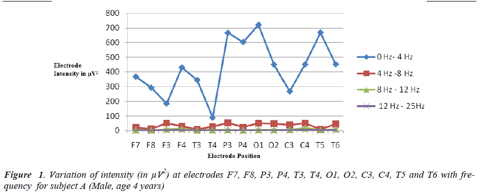
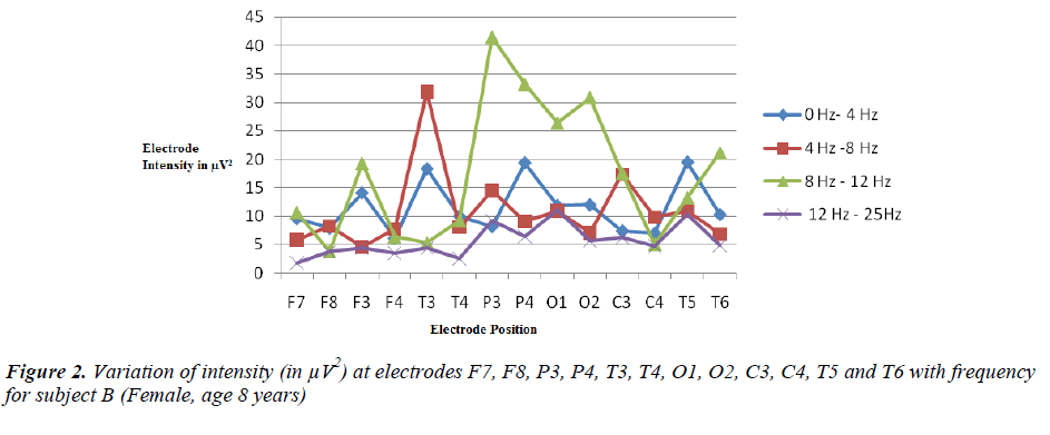
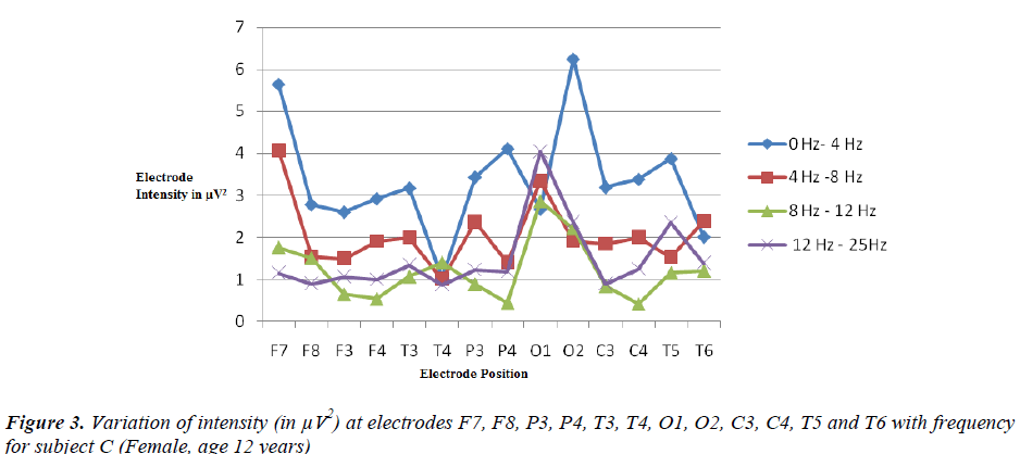
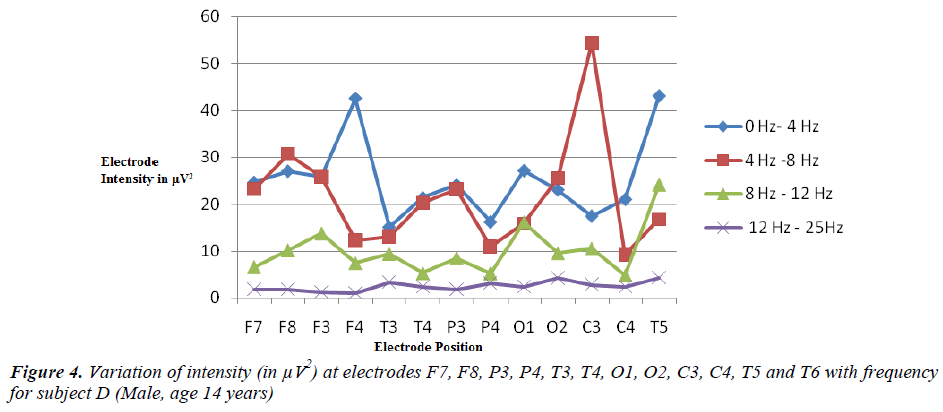
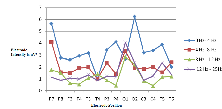
The intensity was measured at electrodes F7, F8, P3, P4, T3, T4, O1, O2, C3, C4, T5 and T6 in four frequency bands as shown below:
Results and Discussion
From the graphical frequency analysis of subjects A, B, C and D, it was observed that the brain activity is different for each subject irrespective of age and gender. The intensities of the signals recorded at electrodes placed near left hemisphere and right hemisphere of the brain were not equal. In the frequency band 0 Hz to 4 Hz, the intensity of EEG signal recorded at all the electrodes was found to be the maximum for the youngest subject A. Maximum intensity for subject A was recorded at electrode O1 and minimum at T4. Electrode O1 is located near the brain activity centre responsible for primary visual while T4 near the centres for emotional processing. The relation between the intensity at the electrode T4 with subjects of different age groups and its effect on emotional development of the subjects could be an interesting research study. In the frequency band 0 Hz to 4 Hz the intensity difference in the similar electrodes were found to be much larger between subjects A & B, A & C and between A and D. The intensity difference was not found much between subjects B, C and D. It was also observed that all the subjects showed more intensity at T5 than at T6. T5 and T6 are localized near the centres of brain activities which are responsible for certain memory functions. In the frequency band 4 Hz to 8 Hz, youngest subject A showed maximum intensity at F3, F4, P3, P4, O1, O2, C4 and T6. Subject A records maximum power at C4 which is located near the centres for sensory and motor functions in the brain. Not much difference at difference electrodes was observed for subjects B, C and D in this frequency band. Subject A recorded least intensity at F7, F8, O1, O2 and T5 in the frequency band 12 Hz to 25 Hz. Subject D, who was the oldest among all, recorded least intensity at F7, F3, F4, T3, T4, P3, P4, C3, C4 and T6. From the above results it is inferred that all the four subjects have unique brain wave patterns. Subject A shows high brain wave activity in the frequency range 0 Hz to 4 Hz while subject B shows maximum intensity at P4 in frequency band 8Hz –12 Hz which suggests better ability of perception and differentiation in subject B. This subject also shows high intensity at T3 in 4 Hz to 8 Hz frequency bands. T3 is located near the centres of emotional processing in the brain. Subject C records high intensity at C3 suggesting better sensory and motor functions. Subject D shows high intensities at O2, O1, F7, and P3 in frequency band 4 Hz to 8 Hz. F7 is located near the centres of rational activities in the brain.
Conclusion
From the present study it is inferred that various regions of the brain do not emit the waves of same frequency when a visual stimulus is administered. The brain waves emitted from various regions have specific effect on the behavior and mental state of a person. There were huge variations found in the brain activity of left and right hemisphere. The large variations in hemispheric activity may be partly due to individual variation in patterns of asymmetric parietemporal hemispheric activity [8]. An EEG signal between electrodes placed on the scalp consisted of many waves with different characteristics. The brainwave patterns were found to be unique for individual subjects. This study suggests that it is possible to distinguish persons according to their typical brain activity. The behavior of a person can also be predicted by analysis of his brain activity. A rational type of person may show increased activity in his frontal left and right hemispheres. From the graphical frequency analysis, it is observed that the brain activity was observed maximum for the youngest subject in frequency band 0 Hz to 4 Hz . It suggests that delta activity is dominant for subject A (age 4 years). Delta waves are marked by an unconscious state, and the loss of physical awareness as well as the "iteration of information". Delta wave activity has also been purported to aid in the formation of declarative and explicit memory formation [9]. Although delta and theta rhythms are generally prominent during sleep, but in present study, delta rhythms are recorded from subject who was awake.
This study also indicates that the brain waves emitted from the left hemisphere are different from that emitted from right hemisphere and therefore the regions of the brain in both the hemisphere show different brain activity and perform different functions. The future research aims to study the importance of the functional asymmetry of the brain for the organization of emotions though the relative contribution of the left and right hemispheres. It also aims to find a relation between the intensity at a particular electrode for the subjects of same age and their emotional response to a given stimuli. The results of such studies can be utilized for developing brain-computer interface applications which will be of great use to physically challenged people.
References
- Xizheng Z, Yin ling, Wang W. “Wavelet Timefrequency Analysis of Electro-encephalogram (EEG) Processing,” International Journal of Advanced Computer Science and Applications, 2010; 1(5): 1-5.
- Aine CJ. A conceptual overview and critique of functional neuro-imaging techniques in humans: I. MRI/fMRI and PET. Critical Reviews in Neurobiology 1995; 9 (2-3): 229-309.
- AlMejrad AS. Human Emotions Detection using Brain Wave Signals :A challenging, European Journal of Scientific Research 2010; 44 (4): 640-659.
- Teplan M. Fundamentals of EEG Measurement’, Measurement Science Review 2002; 2 (2): 1-11. Nunez L., Neocortical Dynamics and Human EEG Rhythms, Oxford University Press, New York.
- Nuwer MR. “Paperless Electroencephalography.” Sem Neurol 1990; 10; 156-165.
- Nuwer MR. Assessment of digital EEG, Quantitative EEG and EEG brain mapping;” report of the American Academy of Neurology and American Clinical Neurophysiology Society. Neurol 1997; 49: 277- 292.
- Levy J, Heller W, Banich MT, Burton LA. Are variations among right- handed individuals in perceptual asymmetries caused by characteristic arousal differences between the hemispheres? J Exp Psychol Hum. Percept. Perform. 1983; 9: 329-358.
- Hobson J, Pace-Schott E. The Cognitive Neuroscience of Sleep: Neuronal Systems, Consciousness and Learning, Nature Reviews Neuroscience 2002; 3(9): 679-693.


