ISSN: 0970-938X (Print) | 0976-1683 (Electronic)
Biomedical Research
An International Journal of Medical Sciences
Research Article - Biomedical Research (2017) Volume 28, Issue 9
Role of Chloride Channel-3 (ClC-3) in SIN-1-induced apoptosis in rat hippocampal neurons
Objective: This study was to investigate the role of Chloride Channel-3 (ClC-3) in apoptosis of primary rat hippocampal neurons.
Methods: SIN-1 (3-morpholinosyndomine), a nitric oxide donor, was used to induce apoptosis in the hippocampal neurons. The cell viability was assessed by the MTT (3-(4, 5-Dimethythiazol-2 yl)- diphenytetra zolium bromide) assay. Hoechst 33342 staining was performed to detect apoptosis. Immunofluorescence staining, Western blot and quantitative real-time PCR were used to evaluate protein and mRNA expression levels.
Results: MTT assay showed that SIN-1 could decrease the cell viability of rat hippocampal neurons, which could be restored by the chloride channel inhibitor, DIDS (4, 4’-diisothiocyanostilbene-2, 2’- disulfonic acid). Results from the immunofluorescence and Hoechst 33342 staining showed that the expression level of ClC-3 was dramatically elevated, and the apoptosis rate was significantly increased, in the SIN-1 group. However, the DIDS treatment could decrease the expression levels of ClC-3, and inhibited SIN-1-induced apoptosis of neurons. Moreover, Western blot analysis showed that the levels of cleaved caspase-3 were significantly elevated following SIN-1 induction, which could be inhibited by the DIDS treatment.
Conclusion: ClC-3 is involved in SIN-1-induced apoptosis in rat hippocampal neurons. These findings might provide theoretical and experimental evidence for the development and application of antiapoptotic drugs in clinical practice.
Keywords
Chloride channel-3 (ClC-3), SIN-1, DIDS, Hippocampal neurons, Apoptosis.
Introduction
Apoptosis occurs under a variety of physiological and/or pathological conditions in diverse cell types, in which multiple pathways might be involved [1,2]. It has been widely accepted that apoptosis affects disease progression, including cancer, chronic inflammation, myocardial infarction, and stroke [3]. Similar characteristic alterations can be observed in various apoptotic cells, including cellular shrinkage, nuclear condensation, DNA fragmentation, and formation of apoptotic bodies [4]. In addition, cell volume loss has been considered as a common feature in virtually all kinds of cellular apoptotic models [5,6]. Recent studies have provided compelling evidence that chloride channels underlie the loss of cell volume in apoptotic pathology, which is closely linked with a series of apoptotic events in different cell types [7,8]. These channels play important roles in the regulation of chloride homeostasis and cell volume under pathophysiological conditions [9]. Moreover, blockers of chloride channels have been shown to possess protective effects against apoptotic responses [8,10]. Among the chloride channel family members, Chloride Channel-3 (ClC-3) is believed to be located on the plasma membrane of mammalian cells, which is associated with the alterations in cell volume, and subsequently involved in the regulation of cell migration, proliferation, differentiation, and apoptosis [11-13]. However, whether ClC-3 could regulate neuronal apoptosis and the underlying mechanism(s) still remain to be established. In this study, SIN-1 (3- morpholinosyndomine), a nitric oxide donor, was used to induce apoptosis in rat hippocampal neurons, and the role of ClC-3 in apoptosis was investigated. Our results showed that ClC-3 expression level was elevated in hippocampal neuronal apoptosis induced by SIN-1, which might be involved in the regulation of the apoptotic process.
Materials and Methods
Primary rat hippocampal neuron culture and grouping
New-born Sprague-Dawley (SD) rats within 1 d after birth, males or females, were provided by the Experimental Animal Center of Guangdong Province (Guangzhou, Guangdong, China). All animal experiments were approved by the Experimental Animal Ethics Committee of Guangdong Province, and complied with the Guide for the Care and Use of Laboratory Animals [14].
Primary rat hippocampal neurons were isolated and cultured as previously described [15,16]. Briefly, new-born SD rats were quickly decapitated, and the brains were removed under sterile conditions. Bilateral hippocampi were separated in cold DHank’s solution and cut into 1 mm3 pieces, followed by digestion with 0.25% (w/v) trypsin (Gibco, Carlsbad, CA, USA) at 37°C for 15 min. Neurobasal medium was added to terminate the digestion, and cells were dispersed uniformly and filtered a 400 mesh filter. Suspension was centrifuged at room temperature at 1000 rpm for 10 min. Cells were then suspended and seeded on culture plates pre-coated with polylysine (Sigma, St. Louis, MO, USA) at a density of 4 × 10⁵ cells/ml, and cultured in a 37°C, 5% CO2 humidified incubator. Culture medium contained 90% (v/v) neurobasal medium, supplemented with 10% (v/v) bovine serum, 2% (v/v) B27, 10 UI/ml penicillin and streptomycin (all from Gibco). 48 h later, the cell culture was subjected to the treatment of 1 × 10⁻⁵ mol/L cytosine arabinofuranoside (Sigma) for 48 h to inhibit glial cell proliferation. Neurons after another 12 d culture were used for experiments, and the cells were identified using immunofluorescence staining with anti-NeuN antibody (Sigma).
These cells were divided into the following groups: (1) the control group that was free from intervention; (2) the SIN-1 group that was treated with 0.5 mmol/L SIN-1 (Sigma) for 18 h; and (3) the SIN-1+DIDS ( 4, 4’-diisothiocyanostilbene-2, 2’-disulfonic acid) group that was treated with 0.5 mmol/L SIN-1+0.1 mmol/L DIDS (Sigma) for 18 h. SIN-1 was used to induce apoptosis in the hippocampal neurons and DIDS was used as a chloride channel inhibitor to block CIC-3.
MTT (3-(4, 5-Dimethythiazol-2 yl)-diphenytetra zolium bromide) assay
Cell viability was assessed by the MTT assay. The 20 μl MTT (0.5 mg/ml; Sigma) was added into each well for 4 h incubation. Then the culture medium was discarded, and 150 μl DMSO (Sigma) was added. Optical Density (OD) was detected by a microplate reader. Cell viability was calculated with the following equation: Cell viability (%)=(ODtreatment- ODblank)/(ODcontrol-ODblank) × 100%.
Immunofluorescence and Hoechst 33342 staining
Rat hippocampal neurons cultured for 12 d were fixed with 4% (v/v) paraformaldehyde (Boster, Wuhan, Hubei, China) for 30 min. After washed with PBS (pH 7.4), cells were blocked with goat serum (Boster) at room temperature for 30 min. Then, cells were incubated with rabbit anti-mouse anti-ClC-3 antibody (1:1000 dilution; Gibco) or mouse anti-rat anti-NeuN antibody (1:1000 dilution; Gibco) at 4˚Covernight. Secondary antibody (1:2000 dilution; goat anti-rabbit and goat antimouse, respectively) was used to incubate the cells at room temperature for 1 h. Then cells were subjected to 20 mg/L Hoechst 33342 (Sigma) treatment at room temperature for 15 min. Observation was performed under inverted fluorescence microscope, and the apoptosis rate was calculated.
Quantitative real-time PCRQuantitative real-time PCR
Total RNA was extracted with the Trizol reagent (Invitrogen, Shanghai, China). Real-time PCR was performed with SYBR Green I. ClC-3 specific primers were synthesized by Guangzhou Jetway Biotechnology Co. Ltd (Guangzhou, Guangdong, China). The primer sequences were as follows: ClC-3, forward 5’-CTACTACCACCACGACTG-3’ and reverse 5’-TTGTCACACCACCTAAGC-3’; GAPDH (Glyceraldehyde 3-phosphate dehydrogenase), forward 5’- CTCCCATTCCTCCACCTTTG-3’ and reverse 5’- CCACCACCCTGTTGCTGTAG-3’. PCR cycling conditions were: denaturation at 93°C for 2 min, then 40 cycles of 93°C for 30 s, 55°C for 45 s, and 72°C for 30 s. Products were detected by 1.0% agarose gel electrophoresis, and the results were analysed using an image analysis system (ABI PRISM 7000 Sequence Detection System, Life Technologies, Shanghai, China). GAPDH was used as control reference.
Western blot analysis
Neurons were harvested and lysed with lysis buffer on ice for 30 min, and then centrifuged at 12000 Xg at 4°C for 15 min. The supernatant was collected, and the protein concentration was determined with a BCA kit (Beyotime, Haimen, Jiangsu, China). Equal amount of protein from each group was subjected to SDS-PAGE. Then the protein was electrophoretically transferred onto a polyvinylidene fluoride membrane. The membrane was blocked with 5% (w/v) defatted milk at 4˚C overnight, and then incubated with rabbit anti-mouse anti-caspase-3 polyclonal antibody (1:1000 dilution; Gibco) and rabbit anti-mouse anti-ClC-3 polyclonal antibody (1:800 dilution; Gibco), respectively, at 4°C overnight. After rinsed with Tris-buffered solution (8.0 g NaCl, 6.0 g Tris, and 1.0% Tweeen-20), the membrane was incubated with horseradish peroxidase-conjugated goat anti-rabbit secondary antibody (1:5000 dilution; Gibco) at room temperature for 1 h. Exposure with conducted with enhanced chemiluminescence reagent. The optical densities of bands were quantitatively analysed with the ImageJ software. GAPDH was used as control reference.
Statistical analysis
Data were expressed as mean ± SD. SPSS13.0 software was used for statistical analysis. One way variance analysis (ANONA) and t-test were applied for the comparisons between groups. P<0.05 was considered statistically significant.
Results
SIN-1 induces neuronal viability decline
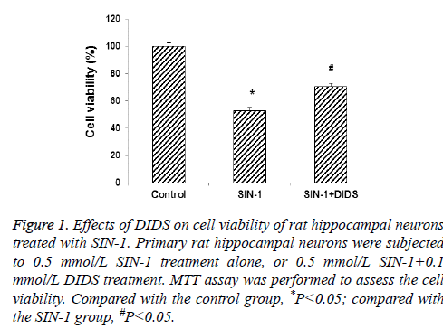
To investigate the cell viability of rat hippocampal neurons following SIN-1 and DIDS treatments, the MTT assay was performed. Primary rat hippocampal neurons were subjected to 0.5 mmol/L SIN-1 treatment alone, or 0.5 mmol/L SIN-1+0.1 mmol/L DIDS treatment. As shown in Figure 1, the MTT assay indicated that, compared with the control group, SIN-1 treatment significantly decreased the cell viability in rat hippocampal neurons (53.41 ± 1.26%) (P<0.05). However, the DIDS treatment significantly restored the decreased cell viability induced by SIN-1 in these cells (69.83 ± 1.13%) (P<0.05).
Figure 1: Effects of DIDS on cell viability of rat hippocampal neurons treated with SIN-1. Primary rat hippocampal neurons were subjected to 0.5 mmol/L SIN-1 treatment alone, or 0.5 mmol/L SIN-1+0.1 mmol/L DIDS treatment. MTT assay was performed to assess the cell viability. Compared with the control group, *P<0.05; compared with the SIN-1 group, #P<0.05.
SIN-1-induces apoptosis in rat hippocampal neurons
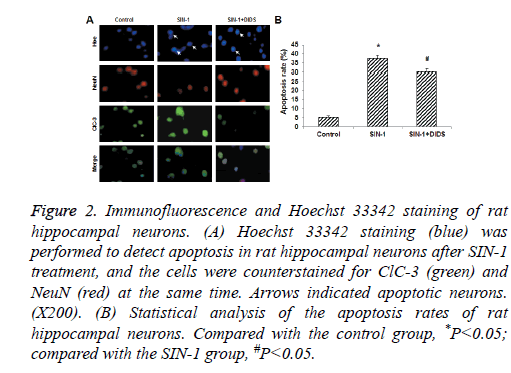
Hoechst 33342 staining was next performed to detect apoptosis in rat hippocampal neurons with SIN-1 treatment. To investigate the involvement of ClC-3 in SIN-1-induced apoptosis, these neurons were counterstained for ClC-3 and NeuN at the same time. Immunofluorescence and Hoechst 33342 staining indicated that ClC-3 was expressed in the rat hippocampal neurons (Figure 2A). Our results showed obvious apoptotic alterations in the SIN-1 group, and the expression level of ClC-3 was dramatically elevated following SIN-1 treatment. Statistical analysis showed that, compared with the control group, the apoptosis rate was significantly increased in the SIN-1 group (Figure 2B) (P<0.05). On the other hand, when these cells were treated with DIDS, the expression of ClC-3 was significantly inhibited, and SIN-1-induced apoptosis was dramatically attenuated. Moreover, the apoptosis rate was significantly elevated compared with the SIN-1 group (Figure 2B) (P<0.05).
Figure 2: Immunofluorescence and Hoechst 33342 staining of rat hippocampal neurons. (A) Hoechst 33342 staining (blue) was performed to detect apoptosis in rat hippocampal neurons after SIN-1 treatment, and the cells were counterstained for ClC-3 (green) and NeuN (red) at the same time. Arrows indicated apoptotic neurons. (X200). (B) Statistical analysis of the apoptosis rates of rat hippocampal neurons. Compared with the control group, *P<0.05; compared with the SIN-1 group, #P<0.05.
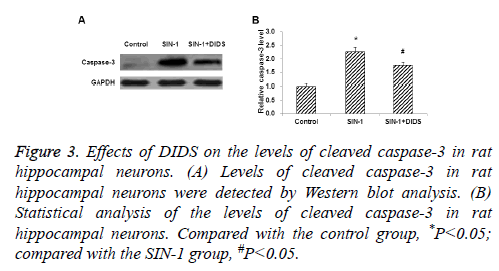
On the other hand, the levels of cleaved caspase-3 were detected by Western blot analysis. Our results showed that the cleaved caspase-3 level was significantly elevated by SIN-1 induction (Figure 3) (P<0.05). As expected, compared with the SIN-1 group, the cleaved caspase-3 level was significantly decreased by DIDS treatment (Figure 3) (P<0.05).
Figure 3: Effects of DIDS on the levels of cleaved caspase-3 in rat hippocampal neurons. (A) Levels of cleaved caspase-3 in rat hippocampal neurons were detected by Western blot analysis. (B) Statistical analysis of the levels of cleaved caspase-3 in rat hippocampal neurons. Compared with the control group, *P<0.05; compared with the SIN-1 group, #P<0.05.
ClC-3 is involved in SIN-1-induced apoptosis in rat hippocampal neurons
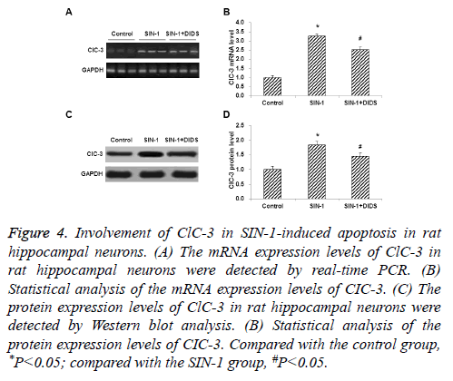
To further investigate the involvement of ClC-3 in SIN-1- induced apoptosis, the mRNA and protein expression levels of ClC-3 in rat hippocampal neurons following drug treatments were detected by real-time PCR and Western blot analysis, respectively. Our results from real-time PCR showed that the mRNA expression level of ClC-3 was significantly elevated in the SIN-1 group, compared with the control group (P<0.05).However, the DIDS treatment dramatically decreased the mRNA expression level of ClC-3 in rat hippocampal neurons (P<0.05) (Figures 4A and 4B). Similar results were obtained with the Western blot analysis. The protein expression level of ClC-3 was increased by SIN-1 administration, which could be decreased by the treatment of DIDS (Figures 4C and 4D) (P<0.05).
Figure 4: Involvement of ClC-3 in SIN-1-induced apoptosis in rat hippocampal neurons. (A) The mRNA expression levels of ClC-3 in rat hippocampal neurons were detected by real-time PCR. (B) Statistical analysis of the mRNA expression levels of CIC-3. (C) The protein expression levels of ClC-3 in rat hippocampal neurons were detected by Western blot analysis. (B) Statistical analysis of the protein expression levels of CIC-3. Compared with the control group, *P<0.05; compared with the SIN-1 group, #P<0.05.
Discussion
Studies show that chloride channels are probably involved in neuronal apoptosis [17]. However, due to the fact that nonspecific chloride channel blockers were used in these previous studies. It is difficult to recognize the specific chloride channel(s) which might be responsible in the apoptotic process. In this study, we investigated the role of ClC-3 in the apoptosis of primary rat hippocampal neurons. SIN-1, a nitric oxide donor, was used to induce neuronal apoptosis in vitro, mimicking the events that occurred in patients with ischemic brain damage. On the other hand, NeuN anti-body is a specific marker for mature neurons, and Hoechst33342 staining is a commonly used method to determine apoptosis [18]. Moreover, caspase-3 plays a key role in cellular apoptosis. Activated caspas-3 fragment (17 kD) is acting as the killer in the apoptotic cascade [19,20]. Our results from the MTT assay, immunofluorescence and Hoechst33342 staining, and Western blot analysis showed that ClC-3 may be involved in the apoptotic process induced by SIN-1 in rat hippocampal neurons, which was in accordance with our previous studies [21].
To further explore whether the ClC-3 was involved in neuronal apoptosis, its mRNA and protein expression levels were detected by real-time PCR and Western blot analysis, respectively. Our results revealed that ClC-3 was expressed in normal neurons, and its expression level was significantly increased in apoptotic cells. Importantly, DIDS could reverse the increased expression of ClC-3 in SIN-1-treated cells. Taken together, the results suggest that ClC-3 may be involved in apoptosis of rat hippocampal neurons induced by SIN-1. Apoptosis is a cascade consisting of multiple signalling pathways. Based on our results and other studies [22], we speculate that caspase-dependent signalling pathway(s) may be involved in SIN-1-induced apoptosis of rat hippocampal neurons. Of course, further in-depth studies are still needed to elucidate the detailed underlying mechanism(s).
In conclusion, our results showed that SIN-1 could decrease the cell viability in rat hippocampal neurons, which could be restored by DIDS. Moreover, the inhibition of ClC-3 attenuated SIN-1-induced apoptosis, suggesting that ClC-3 is involved in SIN-1-induced apoptosis in rat hippocampal neurons. These findings might provide theoretical and experimental evidence for the development and application of anti-apoptotic drugs in clinical practice.
Acknowledgements
This work was supported by the Natural Science Foundation of China (No. 81160157).
Disclosures
All authors declare no financial competing interests. All authors declare no non-financial competing interests.
References
- Cotter TG, Lennon SV, Glynn JG, Martin SJ. Cell death via apoptosis and its relationship to growth, development and differentiation of both tumour and normal cells. Anticancer Res 1990; 10: 1153-1159.
- Poon IK, Lucas CD, Rossi AG, Ravichandran KS. Apoptotic cell clearance: basic biology and therapeutic potential. Nat Rev Immunol 2014; 14: 166-180.
- Lenihan CR, Taylor CT. The impact of hypoxia on cell death pathways. BiochemSoc Trans 2013; 41: 657-663.
- Wyllie AH, Kerr JF, Currie AR. Cell death: the significance of apoptosis. Int Rev Cytol 1980; 68: 251-306.
- Lang F, Ritter M, Gamper N, Huber S, Fillon S. Cell volume in the regulation of cell proliferation and apoptotic cell death. Cell PhysiolBiochem 2000; 10: 417-428.
- Bortner CD, Cidlowski JA. Ion channels and apoptosis in cancer. Philos Trans R SocLond B BiolSci 2014; 369: 20130104.
- Zhang YP, Zhang H, Duan DD. Chloride channels in stroke. ActaPharmacol Sin 2013; 34: 17-23.
- Ko SK, Kim SK, Share A, Lynch VM, Park J, Namkung W, Van Rossom W, Busschaert N, Gale PA, Sessler JL, Shin I. Synthetic ion transporters can induce apoptosis by facilitating chloride anion transport into cells. Nat Chem 2014; 6: 885-892.
- Okada Y, Maeno E, Shimizu T, Manabe K, Mori S. Dual roles of plasmalemmal chloride channels in induction of cell death. Pflugers Arch 2004; 448: 287-295.
- Devuyst O, Guggino WB. Chloride channels in the kidney: lessons learned from knockout animals. Am J Physiol Renal Physiol 2002; 283: F1176-1191.
- Guan YY, Wang GL, Zhou JG. The ClC-3 Cl- channel in cell volume regulation, proliferation and apoptosis in vascular smooth muscle cells. Trends PharmacolSci 2006; 27: 290-296.
- Nilius B, Droogmans G. Amazing chloride channels: an overview. ActaPhysiolScand 2003; 177: 119-147.
- Qian Y, Du YH, Tang YB, Lv XF, Liu J, Zhou JG, Guan YY. ClC-3 chloride channel prevents apoptosis induced by hydrogen peroxide in basilar artery smooth muscle cells through mitochondria dependent pathway. Apoptosis 2011; 16: 468-477.
- The Ministry of Science and technology of the Peoples Republic of China. Guidance Suggestions for the Care and Use of Laboratory Animals 2006; 9-30.
- Daoust A, Saoudi Y, Brocard J, Collomb N, Batandier C. Impact of manganese on primary hippocampal neurons from rodents. Hippocampus 2014; 24: 598-610.
- Li Xi, Yin JB, Zhong ZC, Fan HL, Xu LJ, Zheng YY, Cai SN, Chang QZ. The effects of DIDS on SIN-1-induced rat hippocampal neuronal apoptosis and the expression of PARP/AIF in vitro. Chinese Pharmacological Bulletin 2012; 28: 1549-1552.
- Chang QZ, Zhang SL, Yin JB. Role of chloride channels in nitric oxide-induced rat hippocampal neuronal apoptosis in vitro. Neural Regen Res 2010; 5: 690-694.
- Zhang HN, Zhou JG, Qiu QY, Ren JL, Guan YY. ClC-3 chloride channel prevents apoptosis induced by thapsigargin in PC12 cells. Apoptosis 2006; 11: 327-336.
- Simões PEN, Frozza RL, Hoppe JB, Menezes BM, Salbego CG. Berberine was neuroprotective against an in vitro model of brain ischemia: Survival and apoptosis pathways involved. Brain Res 2014; 1557: 26-33.
- Espinosa-Garcia C, Vigueras-Villasenor RM, Rojas-Castaneda JC, Aguilar-Hernandez A, Monfil T, Cervantes M, Morali G. Post-ischemic administration of progesterone reduces caspase-3 activation and DNA fragmentation in the hippocampus following global cerebral ischemia. NeurosciLett 2013; 550: 98-103.
- Chang QZ, Zhang SL, Yin JB. The role of voltage-dependent chloride channel in rat hippocampal neuronal apoptosis induced by SIN-1. Chin J PharmacolToxicol 2010; 24: 8-11.
- Livak KJ, Schmittgen TD. Analysis of relative gene expression data using real-time quantitative PCR and the 2(-Delta Delta C(T)) Method. Methods 2001; 25: 402-408.


