ISSN: 0970-938X (Print) | 0976-1683 (Electronic)
Biomedical Research
An International Journal of Medical Sciences
- Biomedical Research (2013) Volume 24, Issue 1
Serum markers of tissue damage and oxidative stress in patients with acute myocardial infarction.
1Department of Biochemistry, College of Science, King Saud University, Riyadh, Saudi Arabia
2Division of Clinical Biochemistry, Department of Pathology, Armed Forces Hospital, Riyadh, Saudi Arabia
3Department of Physiology, College of Medicine, King Saud University, Riyadh, Saudi Arabia
4Department of Emergency Medicine, College of Medicine, King Saud University, Riyadh, Saudi Arabia
5Department of Adult Cardiology, Prince Sultan Cardiac Center, Riyadh, Saudi Arabia
- *Corresponding Author:
- Haseeb Ahmad Khan
Department of Biochemistry College of Science
Bldg. 5 King Saud University
P.O. Box 2455, Riyadh 11451
Saudi Arabia
Accepted Date: August 17 2012
Acute myocardial infarction (AMI) is a critical cardiovascular event due to its associated complications and mortality. The pathogenesis of AMI is complex and usually an array of biomarkers is needed for its diagnosis and prognosis. In this study, we determined serum levels of enzymatic markers of tissue damage including aspartate aminotransferase (AST), lactate dehydrogenase (LDH) and creatine kinase (CK) and the markers of oxidative stress including glutathione (GSH) and malondialdehyde (MDA) in 128 AMI patients and 121 normal subjects. We also studied correlations between the above biomarkers and troponin- T, age, gender, smoking and hypertension. The level of LDH was significantly higher in AMI patients (246.49 ± 9.30 U/L) as compared to control subjects (181.63 ± 5.77 U/L). Although the levels of serum AST (29.07 ± 2.06 versus 22.51 ± 0.80 U/L) and CK (133.78 ± 15.23 versus 121.30 ± 11.93 U/L) were higher in AMI patients than controls, they did not reach the significance level. There was a significant decrease in serum GSH and increase in MDA levels in AMI patients as compared to controls. There was no significant correlation between any of the three enzymatic markers of the tissue damage and age indicating their biomarker value irrespective of the age group. However, gender appeared to be a confounding factor while interpreting AST and CK as it showed a significantly negative correlation with the female gender. There were highly significant correlations between troponin-T and AST, LDH or CK. Although smoking was not correlated with any of the biomarkers studied, hypertension showed significant correlation with CK levels. In conclusion, serum LDH is significantly increased in AMI patients that can be regarded as an early predictor of tissue damage. Both the markers of oxidative stress including GSH and MDA are significantly altered in AMI so they can be used to evaluate oxidative stress in these patients. Further studies are warranted to evaluate the prognostic value of these markers in AMI patients.
Keywords
Acute myocardial infarction, Aspartate aminotransferase, Lactate dehydrogenase, Creatine kinase, Glutathione, Malondialdehyde, Oxidative stress, Biomarker
Introduction
Acute myocardial infarction (AMI) is a key component of the burden of cardiovascular disease due to its associated complications and mortality [1]. AMI is the consequence of the chronic development of atherosclerosis lesions. Evidence suggests that reactive oxygen species (ROS) may play important role in the pathogenesis in myocardial infarction [2]. Following ischemia, ROS are produced during reperfusion phase [3,4]. ROS are capable of reacting with unsaturated lipids and of initiating the self perpetuating chain reactions of lipid peroxidation in the membranes [5,6]. In AMI, two distinct types of damage occur to the heart: ischemic injury and reperfusion injury, which lead to mitochondrial dysfunction in heart cells [7]. Diminished antioxidative defense after reperfusion has been found to be associated with impaired myocardial perfusion [8]. Oxidative stress may also lead to the development of serious complications such as left ventricular remodeling after AMI [9] while the redox state of arterial blood can be a predicting factor for left ventricular function after AMI [10].
Aspartate aminotransferase (AST) is an enzyme found mainly in the liver, heart, and muscles. AST is released into the blood by injured liver or muscle cells but is used primarily to detect liver damage. Lactate dehydrogenase (LDH) is an enzyme found in almost all body tissues and plays an important role in cellular respiration. Although LDH is abundant in tissue cells however when tissues are damaged by injury or disease they release more LDH into the bloodstream which can be used to screen for tissue damage. Another enzyme, creatine kinase (CK) is also a better indicator of heart or muscle damage. Assays of serum AST, LDH and CK are widely performed in the early phase of suspected ischemic myocardial injury [11]. CK is a reliable marker for prediction of infarct size and left ventricular function in the acute phase as well as subsequent cardiac events after AMI [12,13]. Murthy and Karmen [14] have found a significant correlation between AST and troponin T in a series of 30 AMI patients.
Although cardiac troponin and CK appear to be the most sensitive and specific markers of myocardial injury there is a need of developing technologies for novel biomarkers or signatures discovery towards point-of-care testing for future management of AMI [15]. Garcia-Pinilla et al [16] have found higher plasma glutathione peroxidase (GPx) activity in patients who experienced acute coronary syndromes and events during follow-up, suggesting GPx as an independent predictor of events during follow-up. Cheng et al [17] have suggested that plasma myeloperoxidase (marker of inflammation) should be considered as a better marker for early diagnosis of AMI. A significant increase in advanced oxidation protein products (AOPP) has been found in AMI patients suggesting its use as a marker of oxidative stress and as a prognostic factor for severe forms of cardiovascular disease [18]. Barsotti et al [19] have observed that advanced oxidation protein products/ thiol ratio may represent a reliable marker of oxidative unbalance in patients with acute coronary syndrome. Lorgis et al [20] validated a new free oxygen radical test (FORT) to assess circulating ROS and concluded that oxidative conditions such as inflammation and diabetes are the major determinants of increased FORT values in patients with AMI. Ho et al [21] have suggested that markers in combination are better for evaluating antioxidant status and monitoring cardiac events than the same markers used separately. In this study, we compared the serum levels of markers of tissue damage (AST, LDH, CK) and markers of oxidative stress including glutathione (GSH) and malondialdehyde (MDA) between AMI patients and control subjects. We also studied correlations between the above biomarkers and the standard cardiac marker (troponin-T) as well as patients’ characteristics including age, gender, smoking and hypertension.
Materials and Methods
We recruited 128 AMI adult patients (91 males, 37 females) admitted to King Khalid University Hospital and Prince Sultan Cardiac Center of the Armed Forces Hospital, Riyadh, Saudi Arabia. Patient exclusion criteria included recent surgery, active infection, chronic inflammatory diseases, diabetes, significant hepatic or renal dysfunction and malignancy. We also included 121 normal subjects for comparative evaluation of various parameters. Peripheral blood samples were obtained from all the patients and controls. Sera were separated for the analysis of enzymatic markers of tissue damage (AST, LDH and CK) and markers of oxidative stress (GSH and MDA). Serum AST, LDH and CK were determined with standard techniques using Cobas 8000 Analyzer (Roche Diagnostics GmbH, Germany). Troponin-T was analyzed using commercially available sandwich ELISA kit (Roche Diagnostics, Germany).
MDA was measured as thiobarbituric acid reactive substances [22]. Serum aliquots were mixed in ice-cold 0.15 M potassium chloride and incubated at 37°C in a metabolic shaker for 1 h. One milliliter of 10% (w/v) trichloroacetic acid was mixed with homogenate followed by centrifugation at 3000 rpm for 10 min. Aliquots (1 mL) of the clear supernatant were mixed with 1 mL of 0.67% (w/v) 2-thiobarbituric acid and placed in a boiling water bath for 10 min, cooled and diluted with 1 mL distilled water. The absorbance of the solution was recorded at 535 nm.
The measurement of GSH in serum was carried out enzymatically according to the modified procedure of Owen [23]. The serum aliquot was mixed with ice-cold perchloric acid (0.2 M) containing 0.01% of EDTA. The homogenates were centrifuged at 10,000 rpm for 5 min. The enzymatic reaction is started by adding 200 μl of clear supernatant in a spectrophotometric cuvette containing 800 μL of 0.3 mM reduced nicotinamide adenine dinucleotide phosphate (NADPH), 100 μL of 6 mM 5,5- dithiobis-2-nitrobenzoic acid (DTNB) and 10 μL of 50 units/ml glutathione reductase (all the above reagents were freshly prepared in phosphate buffer of pH 7.5). The absorbance was measured over a period of 3 min at 412 nm at 30°C.
The data were analyzed by SPSS statistical package (version 10). Mann-Whitney U test (2-tailed) was used to compare means between the patients and controls. Pearson and Spearman correlation tests were used to examine correlations between various biochemical parameters and age, gender, smoking, hypertension, and troponin-T. P values <0.05 were considered as statistically significant.
Results
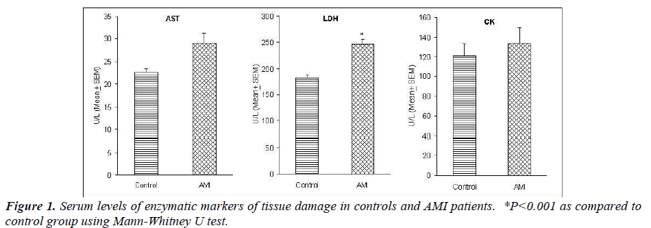
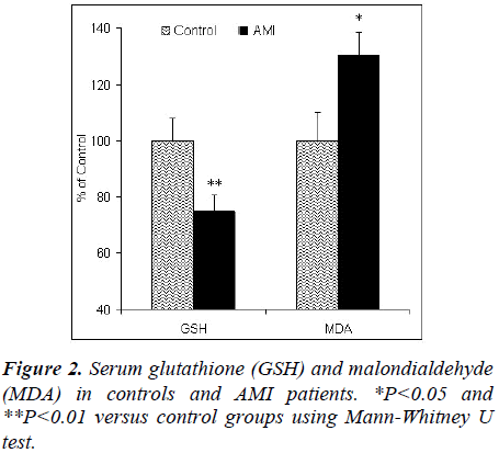
The mean ± SD age of patients and controls were 57.84 ± 12.95 y and 49.01 ± 15.25 y, respectively. The level of serum troponin in AMI patients was 0.783 ± 0.196 ng/ mL whereas all the controls had troponin values of less than 0.003 ng/mL. The level of LDH was significantly higher in AMI patients (246.49 ± 9.30 U/L) as compared to control subjects (181.63 ± 5.77 U/L) (Fig. 1). Although the levels of serum AST (29.07 ± 2.06 versus 22.51 ± 0.80 U/L) and CK (133.78 ± 15.23 versus 121.30 ± 11.93 U/L) were higher in AMI patients than controls, they did not reach the significance level (Fig. 1). There was a significant decrease in serum GSH and increase in serum MDA levels in AMI patients as compared to controls (Fig. 2).
Age was significantly and inversely correlated with GSH (R= -0.172, P=0.034) whereas there was no correlation between age and AST, LDH, CK and MDA (Table 1). A significant correlation was observed between gender and AST (R=-0.162, P=0.012), CK (-0.261, P=0.000) and MDA (R=0.161, P=0.047) however gender was not correlated with LDH and GSH (Table 1). There was no correlation between smoking and any of the biomarkers studied. Hypertension was significantly correlated with CK (R=0.279, P=0.008). There were highly significant correlations between troponin-T and AST (R=0.562, P=0.000), LDH (R=0.589, P=0.000) and CK (R=0.442, P=0.000) (Table 1).
Discussion
Our results showed highly significant correlations between the standard cardiac marker troponin-T and AST, LDH, and CK, indicating potential relevancy of the later markers for predicting cardiac tissue damage, particularly in the small clinics where troponin analysis facility is not available. We observed significantly high levels of serum LDH in AMI patients as compared to controls however there was no significant difference in AST and CK between the two groups (Fig. 1). Experimental study in rats has shown that elevation in the LDH enzyme activity in the serum correlated with a decrease in the activity of cardiac muscle LDH [24]. Swain et al [25] have found that CK:AST ratio is of limited use for the diagnosis of AMI in elderly patients. Gama et al [26] have suggested that measurement of serum CK and AST on the first two days of admission had a sensitivity of 100% and specificity of 86.8% for the diagnosis of AMI; this has been suggested as an optimal combination of cardiac enzymes to achieve maximum sensitivity with the minimum number of tests. The ratio of m-AST to total AST in serum increased after myocardial infarction, being greatest (20%, range 11- 32%) on the third day after onset. For individuals, peak activities of s-AST correlated well with total CK (r = 0.91) and CK-MB (r = 0.86) peak activities, indicating that s-AST also reflects the infarct size [27]. In patients with AMI, measurement of AST provides diagnostic information that differs from that obtained by determination of CK and LDH enzymes and their isoenzymes [27]. Rotenberg et al [28] have suggested that AST should be determined in every patient with suspected AMI, especially in patients admitted 48-72 h after onset of symptoms, when CK declines to near normal values. Cengiz et al [29] have reported a positive correlation between serum LDH and CK levels in AMI patients. Our findings of significantly high levels of LDH and only slight increases in AST and CK suggest that the former is a better indicator of early tissue damage after AMI. There was no significant correlation between any of the three enzymatic markers of the tissue damage and age (Table 1) indicating their biomarker value irrespective of the age group. However, gender appeared to be a confounding factor while interpreting AST and CK as it showed a significantly negative correlation with female gender (Table 1). Although smoking had no significant impact on the levels of these biomarkers, a significant correlation between hypertension and CK could negate the biomarker potential of CK in hypertensive patients.
We observed a significant decrease in serum GSH and increase in MDA in AMI patients (Fig. 2) indicating a state of oxidative stress as reported earlier [30,31]. Another study on 22 AMI patients and 15 controls has found serum GSH levels to be significantly decreased and MDA levels significantly elevated [32]. The GSH/GSSG ratio, indicative of redox status, was found to be lower in AMI patients [21]. The depressed GSH levels may be associated with enhanced protective mechanism to oxidative stress in AMI [32]. Although erythrocyte GSH levels of patients with AMI were significantly depressed (11.5%) as compared to the controls on the second day after infarction, the total mean of GSH levels for all days in patients (3.8% decrease) did not reach statistical significance [33]. While in AMI patients a significant increase in serum MDA was observed in the days following the acute event, reaching a maximum 6-8 days later, when 90% of the patients had values higher than the upper normal limit of the control group [34]. The levels of thiobarbituric acid reactive substances (TBARS, predictor of MDA) were significantly increased and total antioxidant status was significantly decreased in AMI [35,36]. Recently, Bagatini et al [37] have demonstrated a significant increase in TBARS and carbonyl protein levels and a decrease in nonenzymatic antioxidants such as vitamin C and vitamin E levels in 40 AMI patients when compared with the same number of normal subjects. The significantly higher level of MDA in patients with unstable angina and myocardial infarction than in the control group were attributed to significantly higher level of serum cholesterol, high blood pressure, smoking and increased BMI in patients [36]. Intervention with antioxidant Nacetylcysteine has significantly reduced MDA levels in AMI patients [38]. Oral administration of antioxidant Larginine significantly increased the activity of superoxide dismutase, total thiols and plasma ascorbate levels and decreased MDA levels in the sera of AMI patients [39].
Oxidative stress has been regarded as one of the most important contributors to the progression of atherosclerosis [40]. As a natural protective mechanism, myocardial antioxidants inhibit or delay the oxidative damage that consequently prevents thrombosis, myocardial damage and arrhythmias during AMI [41]. However, prolonged oxidative stress due to impaired balance between prooxidant and antioxidant mechanism may lead to lipid peroxidation and tissue damage [42]. There has been a gradual increase in lipid and protein oxidation and a gradual decrease in antioxidant status when the conditions advance from unstable angina to ST-segment elevation myocardial infarction [43]. Blaustein et al [44] have demonstrated that GSH is important in protecting the myocardium against ROS injury and a reduction in cellular GSH content would impair recovery after short periods of ischemia. Experimental studies have shown protective effects of antioxidants against myocardial infarction by inhibiting inflammation and oxidative stress [45-47]. Administration of antioxidants significantly reduced cardiac mortality in patients with AMI [48]. Antioxidant vitamin treatment of AMI patients improves the antioxidant system and reduces oxidative stress, inflammatory process and left ventricular remodeling [49].
In conclusion, serum LDH is significantly increased in AMI patients that can be regarded as an early predictor of tissue damage. Both the markers of oxidative stress including GSH and MDA are significantly altered in AMI so they can be used in tandem to evaluate oxidative stress in these patients. Further studies are warranted to evaluate the prognostic value of these markers in AMI patients.
Acknowledgments
This study was supported by National Plan for Science and Technology (NPST) Program by King Saud University Project Number 08-BIO571-02. We are thankful for the technical assistance of nursing staff of King Khalid University Hospital, Prince Sultan Cardiac Center and Armed Forces Hospital, Riyadh for sample collection and patient care.
References
- Roger VL. Epidemiology of myocardial infarction. Med Clin North Am 2007; 91: 537-552.
- Loeper J, Goy J, Rozenstajin L. Lipid peroxidation and protective enzymes during myocardial infarction. Clin Chim Acta 1991; 196: 119-126.
- Espat NJ, Helton WS. Oxygen free radicals, oxidative stress, and antioxidants in critical illness. Support Line 2000; 22: 11–20.
- Zweier JL, Flahertly JT, Weisfeldt ML. Direct measurement of free radical generation following reperfusion of ischemic myocardium. Proc Natl Acad Sci USA 1987; 84: 1404-1407.
- Slater T. Free-radical mechanism in tissue injury. Biochem J 1984; 222: 1-15.
- Salvemini D, Cuzzocrea S. Therapeutic potential of superoxide dismutase mimetics as therapeutic agents in critical care medicine. Crit Care Med 2003; 31: S29– S38.
- Misra MK, Sarwat M, Bhakuni P, Tuteja R, Tuteja N. Oxidative stress and ischemic myocardial syndromes. Med Sci Monit 2009; 15: RA209-219.
- Kaminski K, Bonda T, Wojtkowska I, Dobrzycki S, Kralisz P, Nowak K, Prokopczuk P, Skrzydlewska E, Kozuch M, Musial WJ. Oxidative stress and antioxidative defense parameters early after reperfusion therapy for acute myocardial infarction. Acute Card Care 2008; 10: 121-126.
- Fujii H, Shimizu M, Ino H, Yamaguchi M, Terai H, Mabuchi H, Michishita I, Genda A. Oxidative stress correlates with left ventricular volume after acute myocardial infarction. Jpn Heart J. 2002; 43: 203-209.
- Ohsawa M, Tsuru R, Hojo Y, Mizuno O, Fukazawa H, Mitsuhashi T, Katsuki T, Shimada K. Relationship between redox state of whole arterial blood glutathione and left ventricular function after acute myocardial infarction. J Cardiol 2004; 44: 141-146.
- Fontes JP, Gonçalves M, Ribeiro VG. Serum markers for ischemic myocardial damage. Rev Port Cardiol 1999; 18: 1129-1136.
- Kazmi KA, Iqbal SP, Bakr A, Iqbal MP. Admission creatine kinase as a prognostic marker in acute myocardial infarction. J Pak Med Assoc 2009; 59: 819-822.
- Mayr A, Mair J, Klug G, Schocke M, Pedarnig K, Trieb T, Pachinger O, Jaschke W, Metzler B. Cardiac troponin T and creatine kinase predict mid-term infarct size and left ventricular function after acute myocardial infarction: a cardiac MR study. J Magn Reson Imaging 2011; 33: 847-854.
- Murthy VV, Karmen A. Troponin-T as a serum marker for myocardial infarction. J Clin Lab Anal. 1997;11:125 8.
- Moe KT, Wong P. Current trends in diagnostic biomarkers of acute coronary syndrome. Ann Acad Med Singapore. 2010;39(3):210-5.
- García-Pinilla JM, Gálvez J, Cabrera-Bueno F, Jiménez- Navarro M, et al. Baseline glutathione peroxidase activity affects prognosis after acute coronary syndromes. Tex Heart Inst J 2008; 35: 262-27.
- Cheng ML, Chen CM, Gu PW, Ho HY, Chiu DT. Elevated levels of myeloperoxidase, white blood cell count and 3-chlorotyrosine in Taiwanese patients with acute myocardial infarction. Clin Biochem 2008; 41: 554- 560.
- Skvarilová M, Bulava A, Stejskal D, Adamovská S, Bartek J. Increased level of advanced oxidation products (AOPP) as a marker of oxidative stress in patients with acute coronary syndrome. Biomed Pap Med Fac Univ Palacky Olomouc Czech Repub 2005; 149: 83-87.
- Barsotti A, Fabbi P, Fedele M, Garibaldi S, Balbi M, Bezante GP, Risso D, Indiveri F, Ghigliotti G, Brunelli C. Role of advanced oxidation protein products and Thiol ratio in patients with acute coronary syndromes. Clin Biochem 2011; 44: 605-611.
- Lorgis L, Zeller M, Dentan G, Sicard P, Richard C, Buffet P, L'Huillier I, Beer JC, Cottin Y, Rochette L, Vergely C. The free oxygen radicals test (FORT) to assess circulating oxidative stress in patients with acute myocardial infarction. Atherosclerosis 2010; 213: 616- 621.
- Ho HY, Cheng ML, Chen CM, Gu PW, Wang YL, Li JM, Chiu DT. Oxidative damage markers and antioxidants in patients with acute myocardial infarction and their clinical significance. Biofactors 2008; 34: 135- 145.
- Utley HG, Bernheim F, Hockstein P. Effect of sulfhydryl reagents on peroxidation in microsomes. Arch Biochem Biophys 1967; 118: 29.
- Owen OG. Determination of glutathione and glutathione disulfide using glutathione reductase and 2- vinylpyridine. Anal Biochem 1980; 106: 207-212.
- Asha S, Radha E. Effect of age and myocardial infarction on serum and heart lactic dehydrogenase. Exp Gerontol 1985; 20: 67-70.
- Swain DG, Gama RM, Nightingale PG. Is serum creatine kinase:aspartate aminotransferase ratio useful for diagnosing acute myocardial infarction in elderly patients? J Clin Pathol 1993; 46: 264-266.
- Gama R, Swain DG, Nightingale PG, Buckley BM. The effective use of cardiac enzymes and electrocardiograms in the diagnosis of acute myocardial infarction in the elderly. Postgrad Med J 1990; 66: 375-377.
- Panteghini M. Aspartate aminotransferase isoenzymes. Clin Biochem 1990; 23: 311-319.
- Rotenberg Z, Weinberger I, Davidson E, Fuchs J, Sperling O, Agmon J. Does determination of serum aspartate aminotransferase contribute to the diagnosis of acute myocardial infarction? Am J Clin Pathol 1989; 91: 91-94.
- Cengiz K, Alvur M, Dindar U. Serum creatine phosphokinase, lactic dehydrogenase, estradiol, progesterone and testosterone levels in male patients with acute myocardial infarction and unstable angina pectoris. Mater Med Pol 1991; 23: 195-198.
- Kabaroglu C, Mutaf I, Boydak B, Ozmen D, Habif S, Erdener D, Parildar Z, Bayindir O. Association between serum paraoxonase activity and oxidative stress in acute coronary syndromes. Acta Cardiol 2004; 59: 606-611.
- Kasap S, Gönenç A, Sener DE, Hisar I. Serum cardiac markers in patients with acute myocardial infarction: oxidative stress, C-reactive protein and N-terminal probrain natriuretic Peptide. J Clin Biochem Nutr 2007; 41: 50-57.
- Kharb S. Low blood glutathione levels in acute myocardial infarction. Indian J Med Sci 2003; 57: 335-337.
- Usal A, Acartürk E, Yüregir GT, Unlükurt I, Demirci C, Kurt HI, Birand A. Decreased glutathione levels in acute myocardial infarction. Jpn Heart J 1996; 37: 177-82.
- Aznar J, Santos MT, Valles J, Sala J. Serum malondialdehyde- like material (MDA-LM) in acute myocardial infarction. J Clin Pathol 1983; 36: 712-715.
- LoPresti R, Catania A, D'Amico T, Montana M, Caruso M, Caimi G. Oxidative stress in young subjects with acute myocardial infarction: evaluation at the initial stage and after 12 months. Clin Appl Thromb Hemost 2008; 14: 421-427.
- Ragab M, Hassan H, Zaytoun T, Refai W, Rocks B, Elsammak M. Evaluation of serum neopterin, highsensitivity C-reactive protein and thiobarbituric acid reactive substances in Egyptian patients with acute coronary syndromes. Exp Clin Cardiol 2005; 10: 250-255.
- Bagatini MD, Martins CC, Battisti V, Gasparetto D, da Rosa CS, Spanevello RM, Ahmed M, Schmatz R, Schetinger MR, Morsch VM. Oxidative stress versus antioxidant defenses in patients with acute myocardial infarction. Heart Vessels 2011; 26(1): 55-63.
- Yesilbursa D, Serdar A, Senturk T, Serdar Z, Sağ S, Cordan J. Effect of N-acetylcysteine on oxidative stress and ventricular function in patients with myocardial infarction. Heart Vessels 2006; 21: 33-37.
- Tripathi P, Misra MK. Therapeutic role of L-arginine on free radical scavenging system in ischemic heart diseases. Indian J Biochem Biophys 2009; 46: 498-502.
- Halliwell B. Free radicals, antioxidants and human disease: curiosity, cause, or consequences? Lancet 1994; 344: 721-724.
- Grech ED, Jackson M, Ramsdale DR. Reperfusion injury after acute myocardial infarction. Br Med J 1995; 310: 477-478.
- Kumari SS, Menon VP. Changes in concentrations of lipid peroxides and activities of superoxide dismutase and catalase in isoproterenol induced myocardial infarction in rats. Ind J Exp Biol 1987; 25: 419-423.
- Serdar Z, Serdar A, Altin A, Eryilmaz U, Albayrak S. The relation between oxidant and antioxidant parameters and severity of acute coronary syndromes. Acta Cardiol 2007; 62: 373-380.
- Blaustein A, Dencke SM, Stolz RI. Myocardial glutathione impairs recovery after short periods of ischaemia. Circulation 1989; 80: 1449-1457.
- Zhou SX, Zhou Y, Zhang YL, Lei J, Wang JF. Antioxidant probucol attenuates myocardial oxidative stress and collagen expressions in post-myocardial infarction rats. J Cardiovasc Pharmacol 2009; 54: 154-162.
- Wang Q, Wang XL, Liu HR, Rose P, Zhu YZ. Protective effects of cysteine analogues on acute myocardial ischemia: novel modulators of endogenous H2S production. Antioxid Redox Signal 2010; 12: 1155-1165.
- Khanna AK, Xu J, Mehra MR. Antioxidant N-acetyl cysteine reverses cigarette smoke-induced myocardial infarction by inhibiting inflammation and oxidative stress in a rat model. Lab Invest 2011 Oct 3. doi: 10. 1038/ labinvest.2011.146. [Epub ahead of print]
- Jaxa-Chamiec T, Bednarz B, Herbaczynska-Cedro K, Maciejewski P, Ceremuzynski L; MIVIT Trial Group. Effects of vitamins C and E on the outcome after acute myocardial infarction in diabetics: a retrospective, hypothesis- generating analysis from the MIVIT study. Cardiology 2009; 112: 219-223.
- Gasparetto C, Malinverno A, Culacciati D, Gritti D, Prosperini PG, Specchia G, Ricevuti G. Antioxidant vitamins reduce oxidative stress and ventricular remodeling in patients with acute myocardial infarction. Int J Immunopathol Pharmacol 2005; 18: 487-496.


