ISSN: 0970-938X (Print) | 0976-1683 (Electronic)
Biomedical Research
An International Journal of Medical Sciences
- Biomedical Research (2006) Volume 17, Issue 2
Short communication: A rare case of vaginal dermoid cyst: A case report and review of literature
Department of Obstetrics and Gynecology, and Reproductive Sciences, St Boniface General Hospital, and University of Manitoba, Winnipeg, Manitoba, Canada
- *Corresponding Author:
- Mesfer Al-Shahrani
Department of Obstetrics and Gynecology, St. Joseph’s Health Care London
268 Grosvener Street, London, ON, Canada N6A 4V2
Phone: 00 1 519 646 6106
Fax: 00 1 519 646 6213
E-mail: mesfersafar ( at ) yahoo.com
Accepted date: May 03 2006
Vaginal dermoid cyst is a rare condition. Ultrasound is the investigative tool for such an entity which may be treated surgically through a transvaginal approach
Keywords
Dermoid cyst, Vaginal cyst
Introduction
The finding of paravaginal dermoid cyst is rare in gynecology practice. The key in the management is the preoperative assessment using transvaginal ultrasound, and the appropriate surgical intervention. We report a case of vaginal dermoid cyst and the surgical procedure in its management.
Case
A 36 year old non-pregnant (G0P0) woman presented to her family physician for treatment refill of her bronchial asthma medications. A gynecological exam had not been performed for the last three and half years. Her family physician decided to perform a well women exam which was difficult to do because of a mass in her vagina.
An ultrasound was requested which showed a solid non-vascular mass measuring 8.4 cm. by 7.5 cm by 6.1 cm, with low level echogenic material as well as echogenic bands, situated within the posterior wall of the vagina, in the rectovaginal space; the rest of the pelvis was normal; with an impression of a dermoid cyst.
The patient was referred to the Gynecology Oncology clinic for further evaluation and management.
She was asymptomatic, and had not been sexually active for the past two years. Prior to that she has been sexually active without any problems. The remainder of her history was non-contributory.
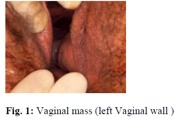
Examination revealed a smooth mass on the left side, just above the vaginal introitus, extending up to the cervix, and was non tender. Pelvirectal exam revealed an appro- ximately 8 by 8 centimeters mass filling the rectovaginal septum to the left.
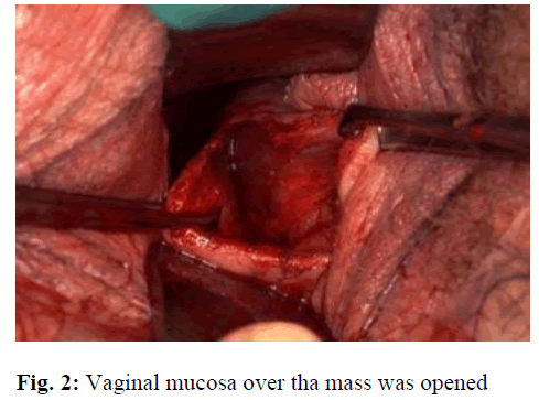
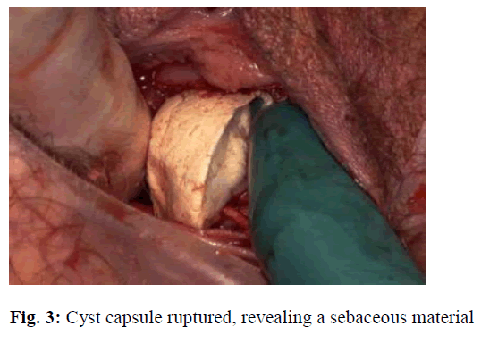
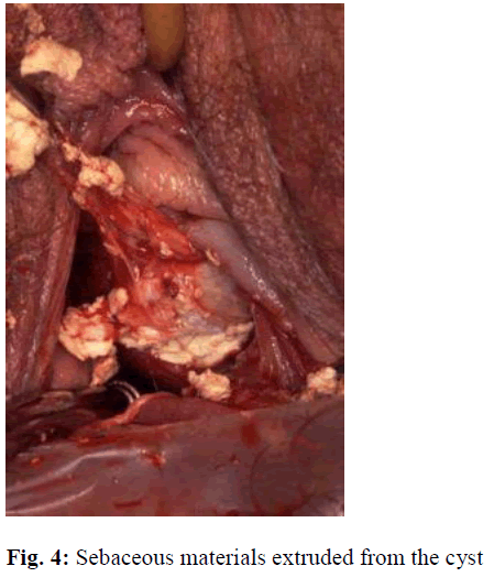
Under general anesthesia this cyst was removed. Incision was made on the vaginal mucosa on top of the mass extended toward the cervix, measuring approximately 6 cm (Figure 1). The mass surface was identified, blunt dissection was carried out (Figure 2), the mass ruptured revealing a sebaceous material (Figures 3, 4) which was extruded and collected in a bowl. The cyst wall was then dissected free from the rectovaginal space with good homeostasis. Incision was sutured with a running locked 0 Vicryl suture, and a vaginal pack was inserted.
The patient was discharged next day after removal of the vaginal pack in good condition. Histopathological examination of the cyst wall, showed keratinized stratified squamous epithelium. There were few sebaceous and apocrine glands noted. The periphery showed chronic inflammation and occasional skeletal muscle tissue. No immature cellular elements were noted. The findings were consistent with a diagnosis of a dermoid cyst.
Discussion
A dermoid cyst (Benign cystic teratoma) is a benign germ cell tumor that contains well-differentiated derivatives of all three germ cell layers.
Ovaries are the commonest site for this tumor, where it is the commonest neoplastic tumor in children and adolescent accounting for more than half of ovarian neoplasms in women younger than 20 years of age. More than 80% of ovarian benign dermoid cysts occur during reproductive years.
A dermoid cyst has also been reported from other parts of the human body other than the ovaries, with rare occurrence. It can be any where in the gastrointestinal tract from the floor of the mouth to the colon [2,3,6,7].
Dermoid cysts have also been reported in male patients, in different parts of their body [6].
Vaginal dermoid is a rare condition. Only five cases have been reported in English literature. First observed in 1899 by Stokes [9], who reported a 44 year old woman who had a 1 centimeter cyst removed from just within the hymen. The cystic contents showed neumerous sebaceous glands and a few hair follicles. Curtis [1] described an ulcerated orange-sized necrotic cyst, containing hair and sebaceous materials in the vaginal mucosa. Johnston [5] described a 4-inch cyst that passed from the vagina in a woman following delivery of her second child. The cyst was filled with thick sebaceous material with matted hair and had been attached to the vaginal wall by a narrow stalk. Hirose et al [4] reported another case, having repeated painful right vaginal wall cyst, which was excised and was found to be a dermoid cyst confirmed by histopatological examination.
Preoperative diagnosis of the exact nature of a vaginal cyst can be difficult. S.S. N. SIU et al [8] reported the sonographic characteristics of a vaginal cyst, which was consistent with a dermoid cyst and was confirmed by histopathological exam after surgical excision of the cyst. The same sonographic features were found in our case.
Transvaginal excision of this type of cyst appears to be an appropriate surgical treatment option which was inconformity with some of the previous cases [4,8,9].
Conclusion
Vaginal dermoid cyst is a rare condition, which can be diagnosed with ultrasound. Transvaginal excision appears to be an appropriate surgical treatment option.
References
- Curtis AH. Transactions of Societies. Chicago Gynecological Society. Surg Gynecol Obstet 1913; 16: 715.
- Fernandez-Cebrian JM, Carda P, Morales V, Galindo J. Dermoid cyst of the pancreas: a rare cystic neoplasm. Hepatogastroenterology 1998; 45: 1874-1876.
- Fujita K, Akiyama N, Ishizaki M, Tanaka S, Ohsawa K, Sugiyama H, et al. Dermoid cyst of the colon. Dig Surg 2001; 18: 335-337.
- Hirose R, Imai A, Kondo H, Itoh K, Tamaya T. A dermoid cyst of the paravaginal space. Arch Gynecol Ob-stet 1991; 249: 39-41.
- Johnston HW. A dermoid cyst of the vagina complicated by pregnancy. Can Med Assoc J 1939; 41: 386.
- Longo F, Maremonti P, Mangone GM, De Maria G, Califano L. Midline (dermoid) cysts of the floor of the mouth: report of 16 cases and review of surgical tech-niques. Plast Reconstr Surg 2003; 112: 1560-1565.
- Schuetz MJ, 3rd, Elsheikh TM. Dermoid cyst (mature cystic teratoma) of the cecum. Histologic and cytologic features with review of the literature. Arch Pathol Lab Med 2002; 126: 97-99.
- Siu SS, Tam WH, To KF, Yuen PM. Is vaginal dermoid cyst a rare occurrence or a misnomer? A case report and review of the literature. Ultrasound Obstet Gynecol 2003; 21: 404-406.
- Stokes JE. The etiology and structure of true vaginal cysts. Johns Hopkins Hosp Rep 1899; 7: 109-136.


