ISSN: 0970-938X (Print) | 0976-1683 (Electronic)
Biomedical Research
An International Journal of Medical Sciences
Research Article - Biomedical Research (2017) Volume 28, Issue 2
Stress analysis of lower back using EMG signal
1Department of Electrical and Instrumentation Engineering, Sant Longowal Institute of Engineering and Technology, Longowal-148106, Punjab, India
2Department of Mechanical, Production, Industrial and Automobile, Engineering, Delhi Technological University, New Delhi, 110 042, India
- *Corresponding Author:
- Pratibha Tyagi
Department of Electrical and Instrumentation Engineering
Sant Longowal Institute of Engineering and Technology
Punjab, India
Accepted date: May 10, 2016
The purpose of this study was to examine the force/EMG relationship during flexion and erection movement of human back during occupation at different positions using surface Electromyography (sEMG) signal. This tool is a non-invasive technique that allows the evaluation of muscle activity. Human’s back is most sensitive part of human body and postures of human body have a significant role to analyze pain especially in the low back region. In this approach surface electrodes are used to record surface electromyography (sEMG) signals of lower back, in the limited forward and backward movement from vertical position, placed at different positions of vertebrae of the lumber region to have a prediction on the stress level of muscles involved in the movement. Preliminary Investigation on three subjects of age groups below 40 years and above 40 years was carried out for three different sitting postures to analyze the differences in EMG signals using Analysis of variance (ANOVA). After Preliminary investigation on three subjects, the experiment was extended to nine subjects in six different sitting postures. ANOVA test has clearly indicated that there exists a statistically significant difference amongst the mean values of EMG signals for different sitting postures and in further investigation, minimum stress level is found in the angle range from 90°-120°. According to the minimum stress level between the angle range 90°-120°, seat may be designed including back rest flexibility in the angle range of 90°-120°.
Keywords
Force, Low back pain, Position of lower back, sEMG.
Introduction
The spine is a complicated structure providing support to the body [1]. One important mechanical function of the lumbar spine is to support the upper body by transmitting compressive and shearing forces to the lower body during the performance of everyday activities [2]. In recent times, low back pain is a common problem in all working professionals. In spite of growing knowledge pertaining to spinal diseases and momentous developments in modern medicine, chronic Low Back Pain (LBP) remains one of the most severe public health problems in all countries including India. Low back pain is the leading musculoskeletal disorder in terms of cost and workabsenteeism [3]. The effectiveness of different kinds of treatments has been studied in the literature, but a definite consensus has yet to be established [4]. LBP causes a socioeconomic impact promoting many days lost in work [5]. Several studies suggest that instability can cause damages and lumbar dysfunctions and increase the risk of an initial episode and subsequent recurrence of LBP [6,7]. Severe back pain most often arises from intervertebral discs, apophyseal joints and sacroiliac joints, and physical disruption of these structures is strongly but variably linked to pain [8]. More of the people with persistent back pain who report limitations in functioning have used health care services compared with others in the sample who also reported functional limitations, presumably resulting from health conditions other than back pain [9]. Therefore, many authors have recommended inclusion in rehabilitation programs of exercises specifically designed to improve active stability of the spine [10,11].
The main motivation of this paper is to study the effect of sitting postures at different angles, which is primarily the main cause of occupational pain. Another purpose of the paper is to investigate the stress level of the muscles involved in these postures using Electromyographic (EMG) signals. Muscle activity is directly reflected by EMG signals. Low muscle activity indicates less energy is required to maintain the posture. So such a study will be useful ergonomic intervention to suggest a proper sitting angle. The proper posture is associated with elastic equilibrium, in which the least elastic stress and lowest joint load are produced [12], which is reflected by the low levels of muscle activity. The proper posture mean the less energy required to maintain the posture and ultimately may result in avoiding occupational health hazard leading to lower back pain. The study embodies the experimental investigation of the physical preparation and data acquisition of the lower back positions. The aspects of data pre-processing stage, which is an essential part to analyze the signal for feature extraction, are also incorporated. Surface electromyography (sEMG) signals are the most common form of non-invasive-measurement of muscle activities [13] and is widely accepted and used for investigation of muscle stress. Extensive researches were made to understand the surface EMG techniques and its application to the analysis of low back muscles for classifying healthy subjects and Low Back Pain (LBP) patients [5].
Materials and Methods
Experimental setup
To improve understanding of the dynamic characteristics of the human lumbar spine, experimental method is required [14]. For this work, MP100 of Biopac System Incorporation has been used for recording EMG signals. MP100 is a complete and expandable data acquisition system that functions like an onscreen chart recorder, oscilloscope and X/Y plotter, which allows recording, viewing, saving and printing data [15].
Data acquisition settings: Muscle activities from the lower back were recorded from the disposable surface electrodes (EL-503) connected to the MP100 Biopac Systems Inc. The data acquisition involves the recording of Electromyographic (EMG) activity [16]. Another important part in data acquisition is the amplification and signal conditioning, which includes artifact elimination of the signals. Since the SEMG signals are relatively small, their measurement is susceptible to the movement of cable that carries signals from the body to the measuring instrument. To eliminate these artifacts, the Electromyogram amplifier module (EMG 100C) high gain, differential input, biopotential amplifier has been used to acquire the EMG with 10-500 Hz bandwidth and gain setting of 2000. The sampling rate was selected to be 1000 Hz so that none of the useful information was lost during data acquisition. The placement procedure of electrodes will be explained in the next subsection.
Placement of electrodes and duration of recording: The surface electrodes were placed with a careful observation of anatomical studies of the muscles concerned with the lower back. EMG data was taken by using two channels of the equipment. The skin preparation was duly done prior to the placement of electrodes. The two active disposable Ag/AgCl surface electrodes were used for each channel in differential configuration at one and half centimeter distance from each other. The third surface electrode was placed as the reference electrode on the unconcerned muscle. Surface electrodes were placed at the skin surface of Erector Spinae at right side and were assigned as Channel 1 for L1 and L3 and Channel 2 for L3 and L5. The placement of channel 1 was to the right side of lumbar vertebrae, L1 and L3 on right erector spine muscle second channel was placed on L3 and L5. All recordings for a subject were taken for each position for a window of 10 sec without back rest.
Subjects and subjects postures
For purpose of the experimental analysis, two stage experiments have been conducted:
Preliminary investigation: Three subjects of age groups below 40 years and above 40 years were considered for three different sitting postures to analyze the differences in EMG signals for a window of 10 sec each.
After Preliminary investigation on three subjects, the experiment was extended to 09 subjects in six different sitting postures without backrest. Healthy subjects (male and female) aged 20-30 years were chosen and they have participated in the experiment with their written consent.
The required essential training for the desired positions of the back was imparted to each subject individually. Back positions were separated by 15 degrees. The six positions of back for which the data was acquired are selected as 45°, 60°, 75°, 90°, 105° and 120° from horizontal plane.
Feature extraction
Generally, most of signals in practice are time-domain signals in their raw format. In other words, one obtains a timeamplitude representation of the signal. The main purpose of the feature extraction is to emphasize the important information in the measured signal. After the successful processing of the sEMG signal, it was required to extract the features of different positions of back. One may easily evaluate the features in time domain because time domain does not need a transformation. Absolute mean and variance time domain features were extracted from acquired EMG signals and has been used for analysis purpose.
Results
For Preliminary investigation, one-way Analysis of Variance (ANOVA), a statistical method was used to test the differences between the mean values of EMG signals of three different sitting postures. The Preliminary test was conducted on three subjects and absolute mean values of muscle activity (EMG) for a window of 10 sec each have been recorded and presented in the Table 1.
| Angle of Back | Subject 1 | Subject 2 | Subject 3 | |||
|---|---|---|---|---|---|---|
| Average Value | Standard Deviation | Average Value | Standard Deviation | Average Value | Standard Deviation | |
| 75° | 0.718743 | 0.563316015 | 0.447931 | 0.12138183 | 1.643806 | 0.9744 |
| 0.703388 | 0.515772916 | 0.417474 | 0.12323651 | 1.708665 | 0.9951 | |
| 0.405779 | 0.281859859 | 0.310476 | 0.12518218 | 1.756806 | 1.0428 | |
| 0.479794 | 0.402271978 | 0.200008 | 0.12300078 | 1.777084 | 1.0467 | |
| 1.485074 | 1.333926257 | 0.264942 | 0.12272625 | 1.775545 | 1.0515 | |
| 0.447859 | 0.410923564 | 0.433159 | 0.12320205 | 1.816283 | 1.0762 | |
| 90° | 0.213493 | 0.155064423 | 0.391967 | 0.266012138 | 0.792189 | 0.4618 |
| 0.212597 | 0.153007881 | 0.327655 | 0.225684525 | 0.845401 | 0.4866 | |
| 0.306643 | 0.397730595 | 0.375299 | 0.247464197 | 0.867605 | 0.4961 | |
| 0.428785 | 0.286482375 | 0.246629 | 0.158845505 | 0.825746 | 0.4885 | |
| 0.471512 | 0.310411638 | 0.333409 | 0.25919276 | 0.900564 | 0.5242 | |
| 0.508391 | 0.35971465 | 0.521975 | 0.337426988 | 0.906899 | 0.5251 | |
| 105° | 0.306535 | 0.148747823 | 0.494207 | 0.11921041 | 0.977155 | 0.5742 |
| 0.243737 | 0.124624189 | 0.499736 | 0.11901171 | 0.985729 | 0.5729 | |
| 0.210168 | 0.113682065 | 0.512191 | 0.11969711 | 0.946091 | 0.5468 | |
| 0.177581 | 0.103052894 | 0.513105 | 0.12025388 | 0.894297 | 0.5216 | |
| 0.154409 | 0.095064539 | 0.509925 | 0.12078698 | 0.8356 | 0.4851 | |
| 0.142599 | 0.089057657 | 0.511921 | 0.12092837 | 0.841688 | 0.4842 | |
Table 1: Feature values of EMG signals for three different angles for preliminary investigation.
Null hypotheses: means of all the EMG signals at different angles of sitting posture are equal.
Alternative hypotheses: means of all the EMG signals at different angles of sitting posture are not equal.
Results of ANOVA test for one subject is presented in Table 2. P-value of ANOVA test for other two were found 0.009447 0.005992 respectively.
| ANOVA: Single Factor | ||||||
|---|---|---|---|---|---|---|
| Subject 3 | ||||||
| SUMMARY | ||||||
| Groups | Count | Sum | Average | Variance | ||
| 75° | 6 | 5.138406 | 0.856401 | 0.001962 | ||
| 90° | 6 | 10.47819 | 1.746365 | 0.003746 | ||
| 105° | 6 | 5.480562 | 0.913427 | 0.004385 | ||
| ANOVA | ||||||
| Source of Variation | SS | df | MS | F | P-value | F critical |
| Between Groups | 2.978146 | 2 | 1.489073 | 442.5829 | 4.61E-14 | 3.68232 |
| Within Groups | 0.050468 | 15 | 0.003365 | |||
| Total | 3.028614 | 17 |
Table 2: ANOVA test summary for three subjects for three different sitting postures.
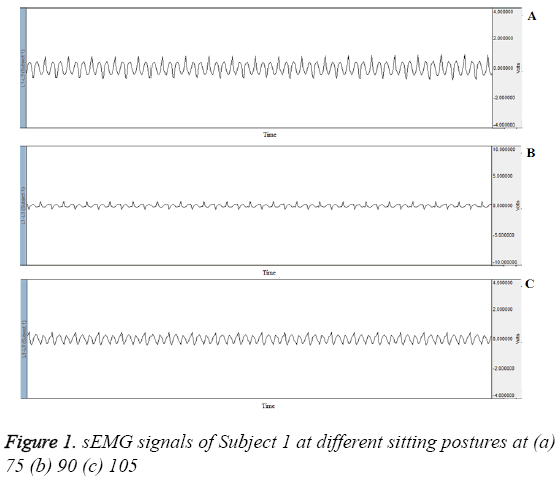
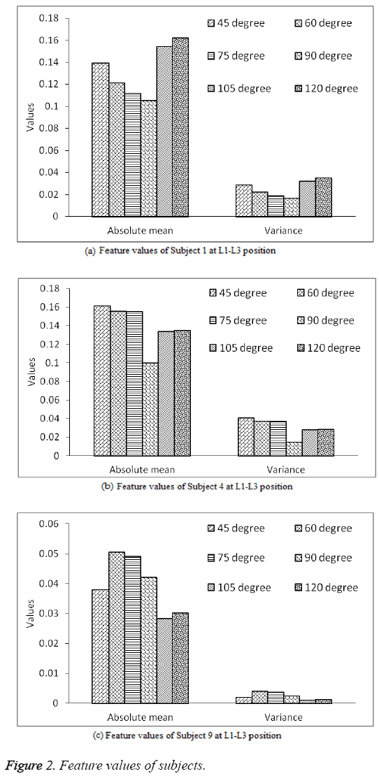
It is clear from the ANOVA method that the p values are considerably lower than 0.05. So the null Hypothesis is rejected and alternate Hypothesis is accepted. It is concluded that the EMG activity is significantly (statistically) different at different angles of sitting posture (Figure 1). Each EMG value represents muscle activity during different sitting with trunk inclinations in flexion and extension positions from the sagittal plane [17]. After the preliminary investigation, the experiment was extended to nine subjects with six different sitting postures without backrest. Two channels for two different locations (L1-L3 and L3-L5) were utilized for each recording. In this analysis, a window of 10 sec. for gross activity and a window of 2 sec. for short duration study have been used for the feature extraction. Figure 2 shows the feature values at six different positions for 10 sec window for three subjects sitting (without backrest) ideally with hands down. It clearly indicates that the EMG output varies for different angle positions of back and comes out to be minimum at 90° in most if the cases.
For further understanding the behaviour of back signals the Table 3 shows the angle for minimum gross stress level of EMG for lower back for the considered nine subjects. It is evident from Table 3, minimum stress level is found maximum times at an angle of 90° i.e. 61%. In rest of the cases the minimum stress level is found at an angle 105° and 120°. Further analysis EMG is analyzed in a smaller window of 2 second each i.e. each 10 sec. recording is divided into 5 parts of 2 sec. each. Table 4 presents the angle of minimum stress level for each 2 second window. It is evident from Table 4, minimum stress level is found maximum times at an angle of 90° i.e. 51% for L1-L3 and L3-L5. Next minimum stress level was found at 105° i.e. 38% for L1-L3 and 11% for L3-L5. Few cases of minimum stress level were found at 120° i.e. 11% and 29% for L1-L3 and L3-L5 respectively. In rest of the cases (9%) the minimum stress level is found at an angle 450 for L3-L5 position.
| Subject | L1-L3 location |
L3-L5 location |
|---|---|---|
| Subject 1 | 105° | 90° |
| Subject 2 | 90° | 90° |
| Subject 3 | 90° | 90° |
| Subject 4 | 90° | 90° |
| Subject 5 | 90° | 120° |
| Subject 6 | 90° | 120° |
| Subject 7 | 120° | 90° |
| Subject 8 | 105° | 105° |
| Subject 9 | 105° | 90° |
| Total | 90°-11 times (61.1%) | |
| 105°-4 times (22.2%) | ||
| 120°-3 times (16.6%) | ||
Table 3: Angle for minimum gross stress level of EMG.
| Subject | L1-L3 location | L3-L5 location | ||||||||||
|---|---|---|---|---|---|---|---|---|---|---|---|---|
| 45° | 60° | 75° | 90° | 105° | 120° | 45° | 60° | 75° | 90° | 105° | 120° | |
| 1 | 0 | 0 | 0 | 0 | 5 | 0 | 0 | 0 | 0 | 5 | 0 | 0 |
| 2 | 0 | 0 | 0 | 5 | 0 | 0 | 0 | 0 | 0 | 5 | 0 | 0 |
| 3 | 0 | 0 | 0 | 5 | 0 | 0 | 0 | 0 | 0 | 5 | 0 | 0 |
| 4 | 0 | 0 | 0 | 2 | 3 | 0 | 0 | 0 | 0 | 2 | 0 | 3 |
| 5 | 0 | 0 | 0 | 5 | 0 | 0 | 0 | 0 | 0 | 0 | 0 | 5 |
| 6 | 0 | 0 | 0 | 5 | 0 | 0 | 0 | 0 | 0 | 0 | 0 | 5 |
| 7 | 0 | 0 | 0 | 0 | 0 | 5 | 0 | 0 | 0 | 5 | 0 | 0 |
| 8 | 0 | 0 | 0 | 1 | 4 | 0 | 0 | 0 | 0 | 0 | 5 | 0 |
| 9 | 0 | 0 | 0 | 0 | 5 | 0 | 4 | 0 | 0 | 1 | 0 | 0 |
| Total | 0 | 0 | 0 | 23 | 17 | 5 | 4 | 0 | 0 | 23 | 5 | 13 |
| % of occurrence of min. stress | 0 | 0 | 0 | 51 | 38 | 11 | 9 | 0 | 0 | 51 | 11 | 29 |
Table 4: Minimum stress level of EMG signal at different angles (2 sec window).
Discussion
SEMG has been used in numerous settings to measure voltage output of relative muscle recruitment, in ergonomic analyses when comparing musculoskeletal stress in a specific muscle(s) associated with postures and to evaluate the efficacy of ergonomic interventions [18,19]. The study utilizes the average amplitude measurement from the sEMG to provide quantitative observation of recruitment intensity for specific muscle groups affected by a task. The analysis used average amplitude directly rather than the often-used percent of maximum voluntary contraction, because some subjects had active injuries and were unable to obtain a reliable maximum reading [20].
Mastalerz and Palczewska observed statistical influence of the trunk inclination on erector spinae, gastrocnemius lat. and tibialis anterior (p<0.05) [17]. Similarly, in our preliminary investigation it has been concluded that the EMG activity is significantly (statistically) different at different angles of sitting posture. A study on effect of postural angle on back muscle activity by Kamil and Md Dawal [12] concluded that neutral upper trunk posture, in which the angle deviates between 0° and -5°, minimizes CES and longissimus muscle activation. This posture allows the subject to maximize balance and optimize the proportions of their body mass and framework based on their physical limitations while performing computer tasks. Low muscle activity indicates less energy is required to maintain this posture, because the muscles are at their ideal length in a neutral position. The neutral posture is associated with elastic equilibrium, in which the least elastic stress and lowest joint load are produced [19], which is reflected by the low levels of muscle activity. The neutral upper trunk position can be considered the ideal posture because it encourages proper alignment of the body’s segments such that the least amount of energy is required to maintain a desired position [12]. In our study occurrence of minimum stress is at an angle 900 for 61% of the readings taken (gross EMG) from nine subjects and is obvious that minimum stress level is mostly found in the angle 900 which is equivalent to neutral position.
Conclusion
The positions of back were investigated by the EMG signals. There is difference in feature values of EMG signal for different sitting posture. Further, ANOVA test has clearly indicated that there exists a statistically significant difference amongst the mean values for EMG signals for different sitting postures, which shows the possibility of investigating the good posture of back using EMG signals. The window selected for the analysis helps us to analyze the changes in EMG signals with time, so it is always better to select a proper window before extracting the features. It is also clear from Table 3 and Table 4 that occurrence of minimum stress is at an angle 900 for 61% of the readings taken (gross EMG) from nine subjects and is obvious that minimum stress level is found in the angle range from 90°-120°. This fact was also verified when shorter duration window (2 sec) of EMG was taken for analysis. So the comfort sitting posture maintaining minimum stress of each individual may vary between the angles range of 900-1200 from the horizontal plane. Accordingly, the seat design may include back rest flexibility in the angles range of 90°-120°.
Acknowledgement
The authors wish to express profound gratitude to Director SLIET Longowal for providing them an opportunity to carry out this work in SLIET and sincere thanks are due for HOD (EIE) SLIET, Longowal for providing them the opportunity to carry out this work in Biomedical Research Lab of EIE Department, SLIET Longowal.
References
- Lam SCB, McCane B. Robert Allen Automated tracking in digitized video fluoroscopy sequences for spine kinematic analysis. J Image Vision Computing 2009; 27: 1555-1571.
- Cholewicki J, McGill SM. Mechanical stability of the in vivo lumbar spine: implications for injury and chronic low back pain. Clin Biomechanics 1996; 11: 1-15.
- Bazrgari B, Shirazi-Adl A, Kasra M. Computation of trunk muscle forces, spinal loads and stability in whole-body vibration. J Sound Vibration 2008; 318: 1334-1347.
- Van Tulder MW, Koes B, Malmivaara A. Outcome of non-invasive treatment odalities on back pain: an evidence-based review. European J Spine 2006; 15: S64-S81.
- Cardozo AC, Gonçalves M. Assessment of Low Back Muscle by Surface EMG: In: Applications of EMG in Clinical and Sports Medicine, Intech, 2012.
- Cholewicki J, McGill SM. Lumbar posterior ligament involvement during extremely heavy lifts estimated from, fluoroscopic measurements. J Biomechanics 1992; 25: 17-28.
- Hides JA, Jull GA, Richardson CA. Long-term effects of specific stabilizing exercises for first-episode low back pain. Spine 2001; 26: E243-E248.
- Adams MA, Dolan P. Spine biomechanics. J Biomech 2005; 38: 1972-1983.
- Nagi SZ, Riley LE, L.G. Newby A social epidemiology of back pain in a general population. J Chronic Dis 1973; 26: 769-779.
- O’Sullivan PB. Lumbar segmental “instability”: clinical presentation and specific stabilizing exercise management. Man Ther 2000; 5: 2-12.
- Robison R. The new back school prescription: stabilization training. Part I Occupational Med 1992; 7: 17-31.
- Kamil NS, Dawal SZ. Effect of postural angle on back muscle activities in aging female workers performing computer tasks. J Phys Ther Sci 2015; 27: 1967-1970.
- Ryait HS, Arora AS, Agarwal R. SEMG signal analysis at acupressure points for elbow movement. J Electromyo Kinesiol 2011; 21: 868-876.
- Kasra M, Shirazi A, Drouin G. Dynamics of human lumbar intervertebral joints. Experimental and finite-element investigations. Spine 1992; 17: 93-102.
- https://www.biopac.com/manual/acqknowledge-tutorial/
- Callaghan J P, Gunning LJ, McGill SM. The relationship between lumbar spine load and muscle activity during extensor exercises. Physical Therapy 2016; 78: 8-18.
- Mastalerz A, Palczewska I. The influence of trunk inclination on muscle activity during sitting on forward inclined seats. Acta of Bioeng Biomech 2010; 12: 19-24.
- Aaras A. Relationship between trapezius load and the incidence of musculoskeletal illness in the neck and shoulder. Int J Industrial Ergonomics 1994; 14: 341-348.
- Marras WS. Industrial Electromyography (EMG). Int J Industrial Ergonomics 1990; 6: 89-93.
- Murphey SL, Milkowski AMS. Surface EMG Evaluation of Sonographer Scanning Postures. J Diagnostic Med Sonography 2006; 22: 298-305.

