ISSN: 0970-938X (Print) | 0976-1683 (Electronic)
Biomedical Research
An International Journal of Medical Sciences
Research Article - Biomedical Research (2017) Volume 28, Issue 19
The application of expansive sub-Tenon's block in vitrectomy
Peng Zhang1, Dan Li2*, Jian Zhang1, Jinpeng Chen1, Zhijun Yang2, Juling Lv1, Weiling Wu1 and Shuping Huo1
1Department of Ophthalmology, Ezhou Hospital of Renmin Hospital of Wuhan University, Ezhou Central Hospital, Ezhou, PR China
2Department of Anesthesiology, Ezhou Hospital of Renmin Hospital of Wuhan University, Ezhou Central Hospital, Ezhou, PR China
- *Corresponding Author:
- Dan Li
Department of Anesthesiology
Ezhou Hospital of Renmin Hospital of Wuhan University
Ezhou Central Hospital, PR China
Accepted date: September 14, 2017
Objective: In view of the discomfort caused by sub-Tenon's Block (STB) during vitrectomy, our study aimed to identify a new method of anesthesia that can reduce the pain caused by STB.
Methods: Sixty patients with vitreous opacity, diabetic retinopathy, complicated retinal detachment or lens dislocated into vitreous cavity were selected. Patients were randomly divided in to experimental group and control group (n=30). No significant differences in background information were found between two groups. All patients were treated with vitrectomy. STB was performed for patients in control group, while expansive STB was used in experimental group. Anesthesia and surgery related factors, degree of pain, basic vital signs, anesthesia complications and analgesia grade were recorded and compared between two groups.
Results: Operation time of anesthesia and onset time of anesthesia were significantly shorter in experimental group than in control group (p<0.01), but no significant differences in operation time of surgery, duration of anesthesia and dose of anaesthetic drugs were found between two group. The degree of pain caused by expansive STB was significantly lower than that of STB (p<0.01). Normal heart rate, respiratory rate and blood pressure were observed in both groups of patients during the whole procedure of surgery. No significant differences in anesthesia complications were found between two groups. The analgesic effect of expansive STB was better than that of STB. In addition, no other complications caused by this method were observed during this study.
Conclusion: Compared with STB, expansive STB can shorten the operation time of anesthesia and onset time of anesthesia, and reduce the degree of pain.
Keywords
Sub-Tenon's block, Expansive sub-Tenon's block, Pain, Anesthesia
Introduction
As the most common approach for orbital regional anesthesia in various ophthalmic surgeries, Sub-Tenon’s Block (STB) can provide effective akinesia and anaesthesia to orbit [1]. In addition, numerous clinical studies have proved that the incidence of complications caused by STB that affect sight is much lower than that of needle-based blocks [2]. This technique is firstly introduced into clinical practices by Turnbull in 1884 [3]. After several rounds of modifications in 1990s, STB has become more and more popular worldwide [4,5]. However, the traditional STB is usually performed by making a conjunctival incision using sprung Westcott scissors and blunt forceps at the position 5 to 8 mm away from limbus, and the insertion of a disposable 19-gauge metal cannula into sub-Tenon's space was followed [6,7]. Although this technique can provide higher safety compared with the application of sharp needle [8], conjunctival haemorrhage occurred during this operation can bring severe pain to patients. Therefore, the development of a more effective method of anaesthesia that can reduce pain is always needed.
The insertion of metal cannula into sub-Tenon's space is the main cause of pain in STB. In this study, an incision less STB technique, which is called expansive STB, was developed to treat patients with different types of retinopathy and vitreous lesions. In this newly developed technique, insertion of metal cannula into sub-Tenon's space was not performed, and iris restorer was used to accelerate the spread of anesthetic liquid along scleral surface, besides that, eyeball was pressed lightly to help the spreading of anesthetic liquid. In order to evaluate the application value of expansive STB, the degree of pain and complications of anaesthesia caused by conventional STB and expansive STB in vitrectomy were compared. We found that expansive STB can not only shorten the operation time of anesthesia and onset time of anesthesia, but also reduce the degree of pain. The report is as follows.
Materials and Methods
Patients
A total of 60 patients with vitreous opacity, diabetic retinopathy, complicated retinal detachment or lens dislocated into vitreous cavity were selected in our hospital from January 2015 to January 2017. All patients received vitrectomy. The age of the patients ranged from 18 to 65 y old and the weight ranger from 51 to 75 kg. All patients could understand NRS-11, which is an 11-point scale for patient to report pain by themselves. No history of cardiopulmonary diseases was found in patients and all patients were suitable for vitrectomy. Patients were randomly divided into experimental group and control group, 30 patients in each group. Patients in experimental group were treated with expansive STB, while conventional STB was used in control group. All patients signed informed consent and this study was approved by the Ethics Committee of our hospital.
Methods of anesthesia
Patients in experimental group were treated with expansive STB. Bupivacaine (7.5 g/L) was used for anesthesia. Intubation was not performed to keep the integrity of fascial bursa. Drug spreading was achieved with injection pressure and the assistance of cotton swabs. Oxybuprocaine (4 g/L) was used for topical anesthesia. Eyelids were opened and patients were asked to stare at the lower side of the nose to expose bulbar conjunctiva between religiosus and lateral rectus. Oxybuprocaine (4 g/L) was subcutaneous injected into fascial bursa through the point 8 mm away from limbus girdle. During injection, cotton swabs were used to assist drug spreading to the back of eyeballs by pressing eyeballs. Using the same method, patients were asked to stare at supertemporal region to expose bulbar conjunctiva between musculus rectus and internal rectus muscle. Then oxybuprocaine (4 g/L) was subcutaneous injected into fascial bursa, and cotton swabs were also used to assist drug spreading to the back of eyeballs by pressing eyeballs.
Patients in control group were treated with conventional STB, conjunctival sac was cut and intubation was performed. Lidocaine (20 g/L) and bupivacaine (7.5 g/L) were used for anesthesia, and oxybuprocaine (4 g/L) was used for topical anesthesia. Eyelids were opened and an incision was made on conjunctiva along nasal side and limbus of corneae to expose sclera. A round blunt tube was pushed along sclera to reach equator of eye. Then, fascia anesthesia was performed using the same amount of drug.
Standard for evaluation
The degree of pain was evaluated according to NRS-11, and the 11 points from 0 to 10 were used to represent different degrees of pain. Higher scores represented higher degrees of pain, “0” represented no pain and “10” represented the most severe pain.
Observation indicators
Degree of pain, heart rate, blood pressure and respiratory conditions during operation were recorded. Adverse reactions including nausea, vomiting, constipation, diarrhea, palpitation and dizziness were also recorded. The overall satisfaction on analgesic treatment was also evaluated.
Statistical analysis
SPSS19.0 (SPSS Inc., USA) was used. Normal distribution data were expressed by (͞x ± s), and t-test was used for comparisons between two groups. Non-parametric Mann- Whitney U test was used to process non-normal distribution data. p<0.05 was considered to be statistically significant.
Results
Comparison of general information between two groups
The general information was compared between two groups of patients. There were no significant differences in age, gender, weight, cultural levels, types of disease and course of disease between two groups (Table 1).
| Age | 53.17 (5.84) | 54.87 (5.69) | p>0.05 | |
| Gender | Male | 18 | 17 | |
| Female | 12 | 13 | ||
| Weight | 57.90 (4.85) | 57.53 (4.46) | p>0.05 | |
| Education | Junior high school | 9 | 9 | p>0.05 |
| Senior high school | 11 | 11 | ||
| College degree and above | 10 | 10 | ||
| Type of disease | Vitreous opacity | 12 | 12 | p>0.05 |
| Diabetic retinopathy | 11 | 12 | ||
| Complicated retinal detachment | 4 | 3 | ||
| Lens dislocated into vitreous cavity | 3 | 3 | ||
| Course of disease | 2.01 (0.90) | 2.22 (1.34) | p>0.05 | |
Table 1. Comparison of background information between two groups of patients.
Comparison of anesthesia and surgery related factors between two groups
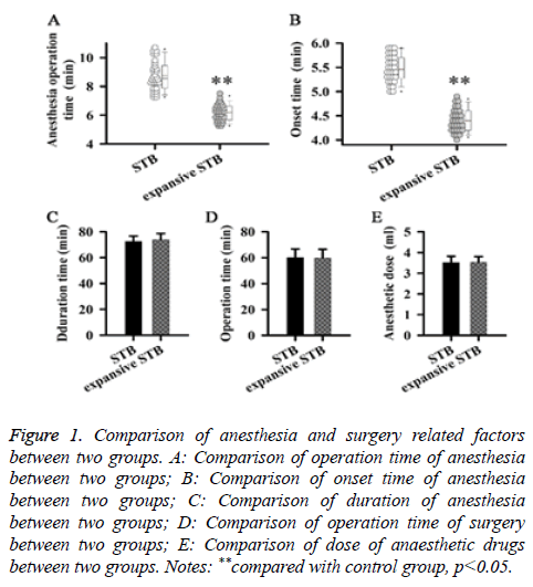
The operation time of anesthesia, onset time of anesthesia, operation time of surgery, duration of anesthesia and dose of anaesthetic drugs were compared between two groups. As shown in Figure 1, the operation time of anesthesia was significantly shorter in experimental group than in control group (p<0.01), in addition, the onset time of anesthesia of experimental group was also significantly shorter than that of control group (p<0.01). No significant differences in operation time of surgery, duration of anesthesia and dose of anaesthetic drugs were found between two groups.
Figure 1: Comparison of anesthesia and surgery related factors between two groups. A: Comparison of operation time of anesthesia between two groups; B: Comparison of onset time of anesthesia between two groups; C: Comparison of duration of anesthesia between two groups; D: Comparison of operation time of surgery between two groups; E: Comparison of dose of anaesthetic drugs between two groups. Notes: **compared with control group, p<0.05.
Comparison of the degree of pain between two groups of patients
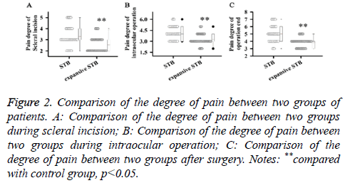
This study aimed to find a better method of anesthesia to reduce the pain of patients. Thus, the degree of pain was compared between two groups of patients. As shown in Figure 2, the degree of pain was significantly lower in experimental group than in control group during scleral incision (p<0.01). In addition, the degrees of pain during intraocular operation and after surgery were both significantly lower in experimental group than in control group (p<0.01). Those results suggest that expansive STB can bring less pain than STB.
Figure 2: Comparison of the degree of pain between two groups of patients. A: Comparison of the degree of pain between two groups during scleral incision; B: Comparison of the degree of pain between two groups during intraocular operation; C: Comparison of the degree of pain between two groups after surgery. Notes: **compared with control group, p<0.05.
Comparison of basic vital signs between two groups of patients
Normal heart rate, respiratory rate and blood pressure were observed during scleral incision, intraocular operation and at the end of surgery. Blood pressure was significantly lower in experimental group than in control group during scleral incision (p<0.01). No significantly differences in other indicators were found between two groups (Table 2).
| Scleral incision | Intraocular operation | Operation end | ||
|---|---|---|---|---|
| HR | STB | 74.27 (6.55) | 73.33 (5.46) | 74.03 (5.22) |
| Expansive STB | 71.87 (6.71) | 70.93 (4.49) | 71.77 (4.79) | |
| RR | STB | 17.30 (1.80) | 16.97 (1.07) | 17.97 (1.38) |
| Expansive STB | 17.50 (1.70) | 17.40 (1.13) | 18.37 (1.43) | |
| BP | STB | 14.80 (1.30) | 13.07 (1.25) | 12.50 (1.14) |
| Expansive STB | 12.77 (1.07)** | 12.53 (1.28) | 12.30 (1.44) | |
Table 2. Comparation of vital signs between two groups.
Comparison of anesthesia complications and analgesia grade between two groups
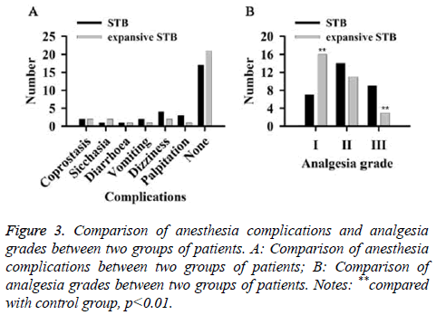
As shown in Figure 3A, the incidence of anesthesia complications was slightly lower in experimental group than in control group, but the differences were not significant. As shown in Figure 3B, the number of patients with analgesia grade I was significantly bigger in experimental group than in control group, while the number of patients with analgesia grade III was significantly smaller in experimental group than in control group. No significant differences in the number of patients with analgesia grade II were found between two groups. Those results suggest that expansive STB can bring better analgesic effect than STB.
Discussion
Numerous clinical studies have proved that STB is a safer and more efficient anaesthetic technique compared with peribulbar anaesthesia [9]. Topical anaesthesia and STB are two most commonly used techniques in cataract surgery. Guay et al. reviewed the available published articles on the application of topical anaesthesia and STB in cataract surgery and they found that both methods are acceptable, but STB is safer and more effective in some cases [10]. In addition, STB was proved to be safe and effective in decreasing additional analgesia and reducing postoperative pain in pediatric strabismus surgery [11]. In another study of strabismus, Ibrahim reported that, compared with IV paracetamol followed by rectal paracetamol, the application of strabismus surgery in children could significantly reduce the incidence rate of postoperative vomiting and agitation [12]. However, the conjunctival incision and the insertion of rigid metal cannula into the sub-Tenon's space in STB can bring pain to the patients. Thus, the development of new approach of anaesthesia in ophthalmic surgeries is always needed. Allman et al. and Lin et al. [13,14] developed a modified STB that can reduce incisions. With that technique, conjunctival incision was avoided by using a 21- gauge, 25 mm, angled and blunt pencil point disposable triport sub-Tenon's cannula. However, the cannula used in this new method is much more expensive than the conventional one, which in turn limited the application of this method. In another study, Kumar et al described another version of STB, within which a reusable cannula can be inserted directly into the sub- Tenon's space without making a conjunctival incision. However, insertion of cannula into sub-Tenon's space is still needed, which inevitably brings pain to patients [1].
In this study, we further modified STB to avoid the insertion of cannula into sub-Tenon's space. We developed expansive STB. With this technique, conjunctival sac was not cut. After surface anesthesia, anesthetic liquid was directly injected into fascial bursa through multi-points. The spreading of anesthetic liquid was assisted using iris restorer. In addition, eyeball was pressed lightly to further improve the spreading of anesthetic liquid. With this operation, the closeness of fascial bursa was kept. Comparison analysis showed that operation time of anesthesia and onset time of anesthesia were significantly shorter in experimental group than in control group. The possible reason is that conjunctival incision and the insertion of cannula into sub-Tenon's space were avoided in this technique, which in turn reduced the operation time of anesthesia. In addition, the methods used in this study can accelerate the spreading of anesthetic liquid, thus shortening the onset time of anesthesia. In this study, no significant differences in operation time of surgery, duration of anesthesia and dose of anaesthetic drugs were found between two groups, indicating that this new technique wound not prolong the operation time and increase the dose of anaesthetic drugs.
Compared with other methods of anesthesia, STB can significantly reduce intraoperative and postoperative pain in various surgical operations [15,16]. However, how to further reduce the degree of intraoperative and postoperative pain is still a hot topic in the field of clinical application of STB. In this study, the degree of pain caused by expansive STB was found to be significantly lower than that of STB, which is possibly due to the avoidance of conjunctival incision and the insertion of cannula into sub-Tenon's space. Normal heart rate, respiratory rate and blood pressure were observed in both groups of patients during the whole procedure of surgery, indicating the high safety of expansive STB. In addition, the overall analgesic effect of expansive STB was also found to be better than that of STB and the incidence of anesthesia related complications caused by expansive STB was also found to be slightly lower than that of STB. Those data suggest that expansive STB is a more effect approach of anesthesia that can bring less pain to patients. It’s also worth to note that this expansive STB avoids the use of the single-use 19-gauge sub- Tenon's cannula, which is approximately 10 USD, thus reducing the economic burden of patients.
In conclusion, Expansive STB can significantly shorten the operation time of anesthesia and onset time of anesthesia. This new technique can also reduce the degree of pain, and inhibit the occurrences of anesthesia-related discomfort or complications. This study is still limited by the sample size. Further clinical studies with bigger sample size are still needed to further confirm the conclusions in this study.
Acknowledgements
We thank the financial support from Science Research Fund of Health and Family Planning Commission of Hubei Province (WJ2015Z102).
References
- Kumar CM, Seet E. Effective and cost-saving incisionless sub-Tenons block. Ind J Anaesth 2017; 61: 84-85.
- Guise P. Sub-Tenons anesthesia: an update. Local Reg Anesth 2012; 5: 35-45.
- Turnbull CS. The hydrochlorate of cocaine, a judicious opinion of its merits. Med Surg Rep 1884; 29: 628-629.
- Hansen EA, Mein CE, Mazzoli R. Ocular anesthesia for cataract surgery: a direct sub-Tenons approach. Ophthal Surg Lasers Imag Retina 1990; 21: 696-699.
- Stevens JD. A new local anesthesia technique for cataract extraction by one quadrant sub-Tenon9s infiltration. Br J Ophthalmol 1992; 76: 670-674.
- Roman SJ, Sit DAC, Boureau CM. Sub-Tenons anaesthesia: an efficient and safe technique. Br J Ophthalmol 1997; 81: 673-676.
- Kumar CM, Dodds C. Sub-Tenons anesthesia. Ophthalmol Clin N Am 2006; 19: 209-219.
- Kumar CM, Eid H, Dodds C. Sub-Tenons anaesthesia: complications and their prevention. Eye 2011; 25: 694-703.
- Roman SJ, Sit DAC, Boureau CM. Sub-Tenons anaesthesia: an efficient and safe technique. Br J Ophthalmol 1997; 81: 673-676.
- Guay J, Sales K. Sub-Tenons anaesthesia versus topical anaesthesia for cataract surgery. Cochrane Lib 2015.
- Tuzcu K, Coskun M, Tuzcu EA. Effectiveness of sub-Tenons block in pediatric strabismus surgery. Braz J Anesthesiol 2015; 65: 349-352.
- Ibrahim AN, Shabana T. Sub-Tenons injection versus paracetamol in pediatric strabismus surgery. Saudi J Anaesth 2017; 11: 72-76.
- Allman KG, Theron AD, Byles DB. A new technique of incision less minimally invasive sub-Tenons anaesthesia. Anaesthesia 2008; 63: 782-783.
- Lin S, Ling RH, Allman KG. Real-time visualisation of anaesthetic fluid localisation following incisionless sub-Tenon block. Eye 2014; 28: 497-498.
- Phillips M, Williamson A, Sivaraj R. Comparison of patient experiences of pain during awake cataract phacoemulsification using sub-tenons block and topical anaesthesia. Anaesthesia 2015; 70: 40.
- Talebnejad MR, Khademi S, Ghani M. The effect of sub-Tenons bupivacaine on oculocardiac reflex during strabismus surgery and postoperative pain: A randomized clinical trial. J Ophthal Vis Res 2017; 12: 296-300.


