Keywords |
| Non-autoimmune thyrotoxicosis, Vitamin D, Pregnancy |
Introduction |
| Gestational transient thyrotoxicosis (GTT) is a condition that is
associated with the stimulation of thyroid stimulating hormone
(TSH) receptor by human chorionic gonadotropin (hCG) [1].
Human chorionic gonadotropin is a glycoprotein hormone. The
similarities between hCG and beta subunit of TSH cause a poor
thyroid stimulant activity. The frequency of hCG-mediated
hyperthyroidism is 1-3% during pregnancy [2-3]. This transient
abnormality arises in the first half of the pregnancy and is
associated with suppressed TSH along with increased T3 and
T4 levels. Graves’ disease can also be diagnosed with a
frequency of 0.1-1% during pregnancy, thus Graves’ disease
should be evaluated in differential diagnosis [4]. |
| It has been reported that serum 25-hydroxyvitamin D level
does not change during the pregnancy but the vitamin D
binding globulin level increases in consistence with estrogen
level. This situation leads to 2-fold increase in 1,25-
dihydroxyvitamin D level. The other causes of the elevated
levels of 1,25-dihydroxy vitamin D are the increased renal 1-
alpha hydroxylase activity and placental 1,25-
dihydroxyvitamin D secretion and release [5-6]. Vitamin D
also has potential benefits on tissues other than skeletal system.
It operates in diabetes mellitus, cardiovascular disease, and
cancer as well as in bone mineralization [7,8-10]. instead of
Vitamin D deficiency is classically known to cause bone mineralization defect but recently, another potential benefits of
vitamin D have gained the importance except the skeletal ones
[7]. Other conditions that come into prominence are the
diabetes mellitus, cardiovascular system and cancer [8-10]. |
| Recently, there are some studies that suggest the low vitamin D
level in pregnancy may be associated with preeclampsia,
preterm delivery and gestational diabetes mellitus [11-12].
There are also some studies that investigate the association
between the thyroid autoimmunity with vitamin D levels. It is
obvious that there is necessity for further studies on this subject
[13]. In our study, we aimed to evaluate thyroid-vitamin D
relationship in GTT. |
Material and Methods |
| We enrolled 83 pregnant women with gestational
thyrotoxicosis and 28 healthy pregnant women, who were
admitted to the Departments of Endocrinology and Obstetrics
and Gynecology in Eskisehir State Hospital, from January
2014 to December 2014. All subjects were within their first
trimester of pregnancy. The diagnosis of thyrotoxicosis was
based on clinical assessment and laboratory findings. All of
patients had thyroid ultrasound. TSH levels with normal or
high serum free T3; free T4 levels were regarded as gestational
thyrotoxicosis. All subjects were tested for TSH-receptor
antibody (TRAb), anti-thyroglobulin antibody (Anti-Tg), anti-thyroid peroxidase antibody (anti-TPO), 25-hydroxyvitamin D,
1,25-dihydroxyvitamin D, calcium and phosphorus. |
| The patients who had positive or borderline positive results of
TSH receptor antibody were excluded from the study. Twentyeight
age and sex matched healthy pregnant women with
normal biochemical and thyroid function tests constituted the
control group. Serum thyroid function tests, thyroid
autoantibodies, erythrocyte sedimentation rate (ESR), vitamin
D, hCG and C-reactive protein (CRP) were measured in all
subjects. The patients did not receive any drug that could
influence the vitamin D level. 25-hydroxyvitamin D levels
below 30 ng/ml were accepted as vitamin D deficiency. |
| Anti-TPO, anti-Tg, TRAb and 1,25-dihydroxyvitamin D were
studied with radioimmunoassay method. 25-hydroxyvitamin D
levels were studied with ELISA method. TSH, free T3 and free
T4 levels were studied with chemiluminescence method. |
Statistical analysis |
| Continues data were given as mean ± standard deviation while
categorical data shown as percentages. Shapiro Wilk test was
used to validate the normality. Paired sample mean test was
used to compare repeated measurements for normally
distributed data; otherwise Wilcoxon Signed Rank test was
performed. IBM SPSS Statistics 21 software was used for
analysis. p<0.05 was assumed to be statistically significant. |
Results |
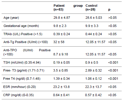
| There was no statistical significance for age, gestational age,
TRAb positivity, anti-Tg positivity, ESR and CRP levels
between the two groups (Table 1). TSH levels were low in
patient group. The control group had higher level of anti-TPO
positivity but when we checked the number of patients who
were positive for anti-TPO, there was no statistical difference. |
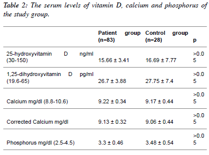
| Additionally there was no statistical difference in vitamin D,
calcium and phosphorus between the two groups (Table 2). But
if we look at carefully to the results we can see the 25-
hydroxyvitamin D levels are below the lower limit in both
groups. On the other hand, 1,25-dihydroxyvitamin D levels of
both groups were within the normal range. |
| Table 3 summarizes the ultrasonographic features. The number
of subjects with thyroid nodules was found more often in
gestational thyrotoxicosis group. The patients with nodules
have one or more nodules. The size of thyroid nodules ranged
between 2 and 30 mm in diameter. As 25-hydroxyvitamin D
levels below 30 ng/ml were accepted as vitamin D deficiency
there is only one patient having 25-hydroxyvitamin D level
over 30 ng/ml [14]. |
| The sizes of the thyroid nodules were with a diameter of
between 2 mm and 30 mm. The presence of nodules was found
to be at risk of 2.67-fold increase for developing gestational
thyrotoxicosis. |
| Table 1: The Characteristics of the Study Population. Anti-Tg:
thyroglobulin antibody, Anti-TPO: thyroid peroxidase antibody, CRP: C-reactive protein levels, ESR: Erythrocyte sedimentation rate, TRAb:
TSH receptor antibody. |
 |
 |
 |
Discussion |
| Gestational transient thyrotoxicosis (GTT) is a condition of
non-autoimmune origin. The stimulation of TSH receptors by
hCG causes hyperthyroidism. This situation becomes more
frequent when the hCG level rises to 70-80000 IU/L levels
[15-18]. In our study we found high hCG levels both in
thyrotoxic and control groups, but the autoantibody levels were
low. |
| In our study we found the hCG levels of the patient group and
the control group were high but the autoantibody levels were
low. The patients with positive TRAb (7 patients) and
borderline levels (6 patients) were excluded from the study to distinguish gestational transient thyrotoxicosis from Graves’
disease. |
| Thyroid function tests of the patients with GTT turned to
normal range during the follow-up. Thyroid ultrasounds were
performed in all participants. One or more nodules were
detected in 42.1% of the GTT patient group and 21.4% of the
control group. Nodules were more frequent in patients with
gestational thyrotoxicosis. The presence of nodules was found
to be 2.67-fold increase for developing gestational
thyrotoxicosis. There are scarcely few studies showing a
relationship between nodules and GTT, but this issue deserves
to be investigated. Vitamin D can cross the placenta and it is
very important for the fetus. Vitamin D deficiency is detected
frequently in pregnancy [19-20]. There are some studies
indicating the importance of vitamin D replacement before and
during pregnancy and after birth [21-22]. The relationship
between Vitamin D and thyroid function has not been not
studied sufficiently. |
| We aimed to evaluate the vitamin D levels in patients with nonautoimmune
gestational thyrotoxicosis in our study. The
vitamin D levels of patients with gestational thyrotoxicosis and
pregnant control group were low and there was no significant
difference between the two groups. It was reported that there
was no relationship between the vitamin D level and thyroid
autoimmunity in one study [13]. Differently, we aimed to
search vitamin D levels in patients with non-autoimmune
thyrotoxicosis etiology. Similarly, we have reached the result
that non autoimmune thyrotoxicosis does not influence the
vitamin D status. We detected low levels of 25-
hydroxyvitamin D, but the levels of 1,25 dihydroxyvitamin D
were within the normal range. This may be due to the
following reasons. First, 25- hydroxyvitamin D is transformed
to 1,25-dihydroxyvitamin D with an increased renal 1 alphahydroxylase
activity during pregnancy. Second, the secretion
and release of placental 1,25-dihydroxyvitamin D increases
during pregnancy (5-6). Calcium and phosphorus levels were
also within the normal range in both groups. |
| Normal levels of calcium and phosphorus which are necessary
for the bone metabolism may be due to normal levels of 1,25-
dihydroxyvitamin D during pregnancy. Normal 1,25-
dihydroxyvitamin D levels in pregnant maintains calcium and
phosphorus balance which is critical in bone metabolism. |
Conclusion |
| Non-auto-immune thyrotoxicosis does not have any effect on
the vitamin D status. The level of 25- hydroxyvitamin D is low
during pregnancy. Preserved level of 1,25-dihydroxyvitamin D
maintains the balanced levels of calcium and phosphorus
which have critical mission in bone metabolism. |
Conflict of Interest |
| The authors declare that there is no conflict of interest. |
References |
- Ballabio M, Poshychinda M, Ekins R. Pregnancy-induced changes in thyroid function: role of human chorionic gonadotropin as putative regulator of maternal thyroid. J Clin Endocrinol Metab 1991; 73: 824-831.
- Krassas GE, Poppe K, Glinoer D. Thyroid function and human reproductive health. Endocr Rev 2010; 31: 702.
- Patil-Sisodia K, Mestman JH. Graves Hyperthyroidism and Pregnancy: A Clinical Update. Endocr Pract 2010; 16: 118.
- Stagnaro-Green A, Abalovich M, Alexander E, Azizi F, Mestman J, Negro R, Nixon A. Guidelines of the American Thyroid Association for the diagnosis and management of thyroid disease during pregnancy and postpartum. Thyroid 2011; 2: 1081-125.
- Glenn D, Braunstein. Endocrine Changes in Pregnancy. In Melmed S, Polonky KS, Larsen PR, Kronenberg HM eds. Williams text book 12. Edition Williams Textbook of Endocrinology 12th Edition. Elsevier Saunders, 2011: 819-832.
- Kovas CS, Fleihan GH. Calcium and bone disorders during pregnancy and lactation. Endocrinol Metab Clin North Am 2006; 35: 2.
- Thacher TD, Clarke BL. Vitamin D insufficiency. Mayo Clin Proc 2011; 86: 50-60.
- Giovannucci E, Liu Y, Rimm EB, Hollis BW, Fuchs CS, Stampfer MJ, Willett WC. Prospective study of predictors of vitamin D status and cancer incidence and mortality in men. J Natl Cancer Inst 2006; 98: 451-459.
- Wang TJ, Pencina MJ, Booth SL. Vitamin D deficiency and risk of cardiovascular disease. Circulation 2008; 117: 503-511.
- Pittas AG, Lau J, Hu FB, Dawson-Hughes B. The role of vitamin D and calcium in type 2 diabetes. A systematic review and metaanalysis. J Clin Endocrinol Metab 2007; 92: 2017-2029.
- Nassar N, Halligan GH, Roberts CL, Morris JM, Ashton AW. Systematic review of first-trimester vitamin D normative levels and outcomes of pregnancy.Am J Obstet Gynecol. 2011; 205: 208.e1-7.
- Urrutia RP, Thorp JM. Vitamin D in pregnancy: current concepts. Curr Opin Obstet Gynecol 2012; 24: 57-64.
- Moon HW, Chung HJ, Hur M, Yun YM. Vitamin D status and thyroid autoimmunity in Korean pregnant women. J Matern Fetal Neonatal Med. 2014: 28: 1-4.
- Bilge U, Ünalacak M, Ünlüoglu I, Ipek M, Çeler Ö, Akalin A. Relationship between 1,25-dihydroxy Vitamin D levels and homeostatic model assessment insulin resistance values in obese subjects. Niger J Clin Pract 2015; 18: 377-380.
- Yoshimura M, Hershman JM. Thyrotropic action of human chorionic gonadotropin. Thyroid. 1995; 5: 425-434.
- Glinoer D, Lemone M.Goiter and pregnancy: a new insight into an old problem. Thyroid 1992; 2: 65-70.
- Glinoer D. Thyroid hyperfunction during pregnancy Thyroid 1998; 8: 859-864.
- Yoshihara A, Noh JY, Mukasa K, Suzuki M, Ohye H, Matsumoto M. Serum human chorionic gonadotropin levels and thyroid hormone levels in gestational transient thyrotoxicosis: Is the serum hCG level useful for differentiating between active Graves' disease and GTT? Endocr J. 2015; 62: 557-560.
- Ginde AA, Sullivan AF, Mansbach JM, Camargo CA Jr. Vitamin D insufficiency in pregnant and non-pregnant women of childbearing age in the United States. Am J Obstet Gynecol 2012; 202: 436-438.
- Sachan A, Gupta R, Das V, Agarwal A, Awasthi PK, Bhatia V. High prevalence of vitamin D deficiency among pregnant women and their newborns in northern India. Am J Clin Nutr 2005; 8: 1060-1064.
- Milman N, Hvas AM, Bergholt T. Vitamin D status during normal pregnancy and postpartum. A longitudinal study in 141 Danish women. J Perinat Med 2011 19; 40: 57-61.
- Li W, Green TJ, Innis SM, Barr SI, Whiting SJ, Shand A, von Dadelszen P. Suboptimal vitamin D levels in pregnant women despite supplement use. Can J Public Health 2011; 102: 308-312.
|


