ISSN: 0970-938X (Print) | 0976-1683 (Electronic)
Biomedical Research
An International Journal of Medical Sciences
Research Article - Biomedical Research (2018) Volume 29, Issue 1
The evaluation of effect of gamma knife on cavernous sinus hemangioma through computed tomography
Min-Xu1, Tao Li2 and Jing Zhang3*
1Imaging Center, Xian Central Hospital, Xian, PR China
2Undergraduate College, Xian No.1 Hospital, Xian, PR China
3Imaging Center, The Women’s and Children’s Hospital of Northwest, Xian, PR China
- *Corresponding Author:
- Jing Zhang
Imaging Center
The Women’s and Children’s Hospital of Northwest Xian, PR China
Accepted date: June 07, 2017
DOI: 10.4066/biomedicalresearch.29-17-997
Visit for more related articles at Biomedical ResearchObjective: Using Computed Tomography (CT) to evaluate the treatment effect of gamma knife surgery in the treatment of Cavernous Sinus Hemangioma (CSHs).
Method: This article retrospectively analysed 31 patients with CSHs who were confirmed clinically and pathologically from Oct 2013 to Apr 2015 in Imaging Center of Xian Central Hospital. Using CT scan images to observe the changes of patient’s clinical symptoms and tumour size before and after gamma knife treatment. The various data were statistically analysed. Further analysis of the influence of tumour lesion size, location and therapeutic dose on the therapeutic effect.
Results: With CT scan, the location of tumour lesion and calcification for CHSs patients can be clearly be seen. After treatment with gamma knife, the diameter of tumour reduced from 3.57 ± 0.46 cm to 2.93 ± 0.56 cm, with significant reduction (P<0.05). From the change of Gross Tumour Volume (GTV) of these patients, 0 cases is complete remission, 22 cases are partial remission, 8 cases are stable and 1 case is progressive. The effective rate of gamma knife is 10.97%, and the stable rate is 96.77%. Furthermore, after treatment with gamma knife, 20 patient’s headache symptoms have been improved, 13 patient’s diplopia symptom have been improved, 5 patient’s tinnitus symptom have been alleviated, 18 patient’s eyelid ptosis symptom have been improved, 21 patient’s facial numbness symptom have been improved. Additionally, the location of tumour lesion is one factor that influences the treatment effect (P<0.05).
Conclusions: Gamma knife treatment could improve nerve function and other symptoms in patients with CHSs, and has a good therapeutic effects and important value.
Keywords
Computed tomography (CT), Cavernous sinus hemangioma (CSHs), Gamma knife treatment
Introduction
The diseases of Cavernous Sinus (CS) are various. Generally include four types such as inflammatory disease, vascular disease, primary tumour and secondary tumours [1]. Compared with other vascular malformations, CSHs is a rare vascular tumour and its incidence is lower. It is a benign, encapsulated and slow-growing space-occupying lesion in cavernous sinus and accounts for 1%-3% of all cavernous sinus tumours. It sees more at middle-aged and older women in their 40’s and 50’s [2,3]. The characteristic of CSHs clinical symptoms is not strong. CSHs has high misdiagnosis rate. It mainly depends on imaging technology to diagnose. Operation resection on the treatment of CSHs is an effective way. The character of its pathology can be defined by surgical management which can reduce the decompression of patient’s important organization and structure caused by tumour. It achieves the purpose of improving clinical symptoms [4]. During primary screening to CSHs patients, CT was widely recognized for its rapid and accurate location, etc. CT plain scan can achieve the preliminary determination of location of tumour lesion and calcification, while enhanced CT scan can show the tumour more clearly [4]. But cavernous sinus has complex structures, adjacent to cranial nerves and is hyper vascular, so it is possible to cause haemorrhage during the procedure, the small probability of whole resection, high probability of cervical node metastasis and so on. It has a relatively high mortality rate and a high rate of morbidity, so there are more risks of operations [5]. Radiosurgery is originally as a complementary medicine which is mainly aimed at remnant carcinoma and old and weak patients. It gets a lot of attention, because of its great treatment effects. It plays an important role in further improving the effect of radiosurgery and reduces the injury during radiosurgery, especially with the emergency with the Gamma knife and cyber knife [6]. This article retrospectively analysed basic clinical information in 31 patients with CSHs who had accepted the gamma knife therapy. Further reviews the changes of patient’s tumour size and clinical symptoms before and after therapy and explore the curative effect and application value of CSHs patients in Gamma knife treatment further from their CT images. From Oct 2013 to Apr 2015 in Imaging Center of Xian Central Hospital, the treatment effect of gamma knife for CSHs was evaluated using CT images, with follow-ups to patients.
Materials and Methods
Clinical information
Retrospectively analyse 31 patients with CSHs confirmed by operation and pathology From Oct 2013 to Apr 2015 in our hospital, aged between 28-63 y and the average age is 42.67 y, 12 were males and 19 were females. The course of disease is 1 month to 3 y. The main clinical symptoms are headache, facial numbness, drooping eyelids and diplopia and so on.
Method of CT scanning
Using 64-slice spiral CT (Light Speed VCT) produced in GE Company to scan, scanning paraments: 120 kV, 250-335 mA, 0.531 pitch, 20-40 cm FOV, 512 × 512 matrix, 5 mm slice thickness. Enhancement scan were performed after the CT plain scan. Through elbow front the intravenous injection of 70 ml Ultravist (370 mg I/ml), 30 ml of normal saline, injection rate are all 4.0 ml/s. The value of enhanced CT scan rises when 20 HU than plain scan which means little enhancement. 20-40 HU is moderate enhancement, more than 40 HU is obvious enhancement.
Gamma knife therapy and dose
Under the premise of local anaesthesia the Leksell stereotactic frame was used. The image of lesions localization can be acquired through CT scanning. The patients are irradiated by using MASEP-SRRS skull gamma ray therapeutic system.
Patients with the tumour measuring more than 4 cm were treated by gamma knife in 2 divided doses by using fractionated dosage and 50% isodose curve. Circumference dose is 8-10 Gy and central dose is 16-20 Gy during the first treatment. Circumference dose is 8-9 Gy and central dose is 16-18 Gy during the second treatment.
Patients with tumour measuring less than 4 cm accept the treatment of single gamma knife and 50% isodose curve. In circumference dose is 9-14 Gy and central dose is 18-26 Gy. All patients after the surgery should review once every six months and the follow-up time is 18-36 months generally.
Varying parameter of tumour
Tumour can be divided into three types according to the maximum length of tumour. Diameter 2.1-3.0 cm for small, 3.1-4.0 cm for medium, 4 cm above is large.
The criteria of tumour volume: Complete Remission (CR) means that target lesion disappear altogether. Partial Remission (PR): The length of the lesion was reduced by more than 50%. Stable Disease (SD): The sum of the length diameter is reduced from 25%-50%; Progressive Disease (PD): The sum of the length and diameter increased by 20% or new lesions appeared. Effective rate=(CR+PR)/Total cases. Index of stability=(CR +PR+SD)/Total cases.
Statistical method
The patients are divided into different groups according to different therapeutic dose, lesion size, lesion location and so on and comparative statistical difference to evaluate the impact of these factors on the prognosis of patients. All data were analysed by SPSS17.0 software. The statistical method: χ2 test is used for enumeration data. T-test is used for comparison between groups. T-test or one-way repeated measures ANOVA is used in comparing. P<0.05 can be considered significant.
Result
CT plain scan and enhanced scan
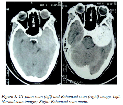
CSHs patient’s CT scan image can find high density lesion (Figure 1). Saddle part exhibit smaller and symmetric “gourd shaped” or “dumbbell”. The tip of lesion grows to sella and juxtaslla. Punctate calcification can be seen in the posterior part. The tumour was equal or slightly high density shadow on one side of the saddle in CT plain scan (Figure 1, left). The margins around the masses are clear. There is edema around the tumour. Obvious and uniform enhancement can be seen in the CT enhanced scan image (Figure 1, right) of the tumour. After treatment, the Gross Tumor Volume (GTV) decreased obviously, as shown in Figure 1.
The change of the tumour before and after the treatment
According to the data in Table 1, the number of small tumours increase and the number of decrease after the treatment. Besides, the tumour diameter is 3.57 ± 0.46 cm before the treatment and The tumour diameter is 2.93 ± 0.56 cm after the treatment which decreased significantly than the before treatment (P=0.029) .
| Tumour diameter (cm) | Before the treatment (n=31) | After the treatment (n=31) | P |
|---|---|---|---|
| 2.1-3.0 (small-scale) | 9 cases | 15 cases | 0.025 |
| 3.1-4.0 (medium-sized) | 10 cases | 9 cases | 1.022 |
| >4.0 (large) | 12 cases | 7 cases | 0.032 |
Table 1. The change of tumour before and after the treatment.
In the view of the change of tumour volume, there is no complete remission. Partial remission is 22 cases. Stable remission is 8 cases. An evolutional case is 1 case. Effective rate is 10.97% and stable rate is 96.77%.
The results show that gamma knife radiosurgery can effectively reduce the size of tumour in patients with CSHs.
Discussion
CSHs come from vascular system of cavernous sinus and are located in cavernous sinus dura mater, which is composed of a high pressure sinus like structure. There is no haemorrhage or cystic change generally. CSH is a rare congenital vascular malformation [7]. CSHs often occur in the middle cranial fossa and cavernous sinus and are more closely related to the dura. It is also known as “Dural cavernous hemangioma”. Most of the CSHs are solitary lesions. The lesions were round or quasi circular, “gourd shaped”, “dumbbell” or irregular leaf division. It grows across the sella and involves the cavernous sinus and sella turcica [8]. Because of its slow growth and a certain growth space, the tumour cannot oppress adjacent important structures and structures before the tumour dose not form a significant position and few clinical symptoms appear [9].
Earlier because the diagnostic means were relatively simple and there were no specific clinical symptoms of CSHs, so it is often misdiagnosed as meningioma or schwannoma [10]. When the patient is diagnosed the tumour lesions were more than 3 cm and accompanied by II-VI cranial nerve dysfunction and cavernous sinus compression such as headache, visual deterioration, diplopia, exophthalmos, ptosis, facial numbness, Abducens nerve paralysis and Oculomotor palsy and so on. Epileptic once was the first symptom of some patients [11]. The disease is more common in women, but there is no genetic predisposition [12].
At present, treatment includes microsurgical resection, embolism, fractionated radiotherapy, stereotactic radiosurgery and so on for symptomatic CSHs. Surgical resection is a common method for the treatment of CSHs. But because of the abundant blood supply of CSHs, bleeding can be massive in operation. Adhesion between tumour and peripheral nerve tissue is very serious. Uncontrollable bleeding and III-VI of cranial nerve injury can occur in operation. The patients lose the function of extra ocular muscle [13] and the total resection rate was lower [14]. Therefore, the risks and complications of the operation cannot be ignored. Much attention also should be drawn to the problem the multiple lesions.
Since 1999, Iwai et al. reported for the first time that 1 patient with residual CSHs was treated with gamma knife radiosurgery and then the lesion was shrinked [15]. When the cranial nerve injury symptoms do not appear, reports on CSHs radiation therapy have appeared one after another, which all showed good therapeutic effect and low complication rate [5]. Some studies indicate that CSHs is sensitive to radiation. Preoperative radiotherapy can reduce tumour size, intratumoral vessel degeneration, stenosis, intratumoral thrombosis, which is advantageous to the operation [16]. With the advent of gamma knife radiosurgery and other therapeutic modalities, radiosurgery from the surgical treatment, gradually become the preferred treatment. Especially for Frail elderly patients and patients with smaller tumours [17]. Based on the observation of the patients with CT scanning image, this paper analyse the changes of clinical symptoms and tumour size in 31 patients with CSHs. The data shows that the diameter of the tumour was changed from 3.57-0.46 cm to+2.93+0.56 cm and tumour diameter was significantly reduced (P<0.05) after gamma knife treatment in CSHs patients. In addition, the effective rate of gamma knife radiosurgery was 10.97%. Stable rate was 96.77%. The results show that gamma knife radiosurgery can significantly improve the tumour lesions in patients with CSHs. This study is consistent with the results of Iwai et al. [15,16]. Furthermore, compared with the situation before treatment, the patient’s chief clinical symptoms, such as headache, facial numbness and eyelid ptosis etc. are better, indicating that their damaged neurological functions been improved to some extents.
After comprehensive analysis of theoretical dose used during gamma knife treatment, influencing factors of tumour lesion size and location, etc. on the prognosis of patients, the single factor analysis shows no significant difference of tumour lesion size, circumference dose and central dose on the prognosis of patients (P>0.05). However, the location of lesion shows significant influence on the therapeutic effect (P<0.05). The multiple-factor analysis, being consistent with the influence on results from single factor analysis, also shows significant difference of lesion location on the therapeutic effect.
Acknowledgement
This research was supported by Fund Project: Science and technology research project of social development in Shaanxi Province (2016SF-180).
References
- Zongjilong LM. Discuss the value of CT and MRI in the diagnosis and differential diagnosis of cavernous sinus cavernous hemangioma. Chinese Med Guide 2011; 9: 132.
- Banyunchao JT, Li T. MRI findings and clinic pathological study of cavernous sinus cavernous hemangioma. Shaanxi Med J 2012; 41: 463-466.
- Yan S. The value of CT and MRI in the diagnosis of cavernous sinus cavernous hemangioma. Chinese J Pract Nervous Dis 2013; 16: 48-49.
- Zhang Y. Comparison study of CT and MRI in the diagnosis of intracranial cavernous hemangioma. Med Innov China 2016; 13: 47-50.
- He Z. Imaging diagnostic features and treatment options for cerebral cavernous hemangioma. J Chinese Neurosurg Dis Res 2016; 15: 378-380.
- Wang X. The role of stereotactic radiosurgery in cavernous sinus hemangiomas: a systematic review and meta-analysis. J Neuro Oncol 2012; 107: 239-245.
- Jin XY, Zhang G. Curative effect observation of gamma knife radiosurgery for cavernous sinus cavernous hemangioma. China Med Herald 2012; 9: 87-88.
- Zhou C, Chen G, Shen G. Long-term efficacy analysis of gamma knife radiosurgery for skull base meningiomas. Chinese J Clin Neurosurg 2011; 16: 117-118.
- Sun S, Liu A, Wang Z. Gamma knife radiosurgery for trigeminal neurinoma. Chinese J Neurosurg 2006; 22: 275-278.
- Du H, Zeng X, Li M. Diagnosis and treatment of intracranial cavernous hemangioma. Shandong Med J 2012; 52: 40-41.
- JaGe ZJ, Ma Z. Analysis of the efficacy of gamma knife radiosurgery for intracranial cavernous hemangioma. J Central South Univ 2014; 39: 1320.
- Mo X, Zhou W, Dong J. Imaging diagnosis and pathologic features analysis of cavernous sinus hemangiomas. J Med Res 2014; 43: 140-144.
- Wang S, Chen H. Diagnosis and treatment of cavernous sinus cavernous hemangioma. Surg Neurol 2012; 8: 832-833.
- Zhong B. Imaging features of cavernous sinus cavernous hemangioma. Int J Neurolog Depart Neurosurg 2013; 40: 5-6.
- Bansal S. Cavernous sinus hemangioma: a fourteen year single institution experience. J Clin Neurosci 2014; 21: 968-974.
- Yang A, Lu J. Clinical value of arterial embolization with lipiodol emulsion in the treatment of giant hepatic cavernous hemangioma in Pingyang. Chinese J Med 2012; 47: 50.
- Qiao N. A systematic review and meta-analysis of surgeries performed for treating deep-seated cerebral cavernous malformations. Br J Neurosurg 2015; 29: 1-7.
