ISSN: 0970-938X (Print) | 0976-1683 (Electronic)
Biomedical Research
An International Journal of Medical Sciences
- Biomedical Research (2016) Volume 27, Issue 2
The intervention effect of the Rhizoma rhizomae t2dm on rat microvascular angina inflammatory-pathway.
| Qi Feng1, Jin Hongguang1, Wang Yiqiang1, Song Baiji2, Deng Yue3* 1The Cardiology Department of Affiliated Hospital of Changchun Traditional Chinese Medicine University, 130000, PR. China 2Changchun, Jilin Changchun city Erdao District Dongsheng community health service center 130000, PR. China 3Heart Disease Center, Jilin Affiliated Hospital of Changchun Traditional Chinese Medicine University 130000, PR. China |
| Corresponding Author: Deng Yue, Heart disease center, Jilin Affiliated Hospital of Changchun Traditional Chinese, Medicine University, PR. China |
| Accepted: January 30, 2016 |
Objective: Observe the intervention effect of the rhizoma Rhizomae t2dm on rat microvascular angina inflammatory-pathway, and carry out the correlation research on coronary blood flow reserve function.
Methods: Micro particle induced by thrombosis for acute thrombosis in coronary microcirculation of rats, according to randomized method, 1 day will divide 60 rats into the experimental group and control group, the blank group, respectively give Rhizoma zedoariae t2dms, nitroglycerin, saline lavage, 28 days after detection of nitric oxide, angiotensin II, thromboxane A2 (TXA2); Monocyte chemotactic protein 1 (MCP 1).
Results: In the aspect of nitric oxide increases, Rhizoma zedoariae t2dm group is better than the nitroglycerin group (P<0.05), and the nitroglycerin group is better than the blank group (P<0.05), so the dilation of coronary vessels can improve coronary blood flow reserve. In the aspect of angiotensin II reduction: zedoary Rhizoma zedoariae t2dm group is better than the nitroglycerin group and the blank group (P<0.05), and the nitroglycerin group has the same therapy effect with the blank group (P>0.05). In the aspect of the thromboxane A2 reduction: Rhizoma zedoariae t2dm group is better than that of nitroglycerin group and blank control group (P<0.05) and the nitroglycerin group has the same therapy effect with the blank group (P>0.05), changing the coronary blood flow due to the reduction blood-vessel shrinking substances. In the aspect of MCP-1 reduction: Rhizoma zedoariae t2dm group is better than the nitroglycerin group and the blank group (P<0.05), and the nitroglycerin group has the same therapy effect with the blank group (P>0.05).
Conclusion: Rhizoma Rhizomae t2dm capsules to improve the function of coronary blood flow reserve.
Keywords |
||||||||
| Rhizoma zedoariae , Microvascular angina pectoris, Inflammatory-pathway, Phlegm and blood stasis fu evil. | ||||||||
Introduction |
||||||||
| Microvascular angina-MVA refers to a group of clinical syndromes with typical angina pectoris symptoms, positive electrocardiogram exercise tests, normal coronary artery angiography, and without coronary artery spasm basis and ruling out other heart diseases able to cause ischemic ECG changes [1]. In 1967, Lidoff at first reported 15 female patients with typical angina pectoris symptoms and ECG positive treadmill tests, but normal coronary artery angiography [2]. In 1973, Kemp named this syndrome as "X syndrome" [2]. Recent research data show that more than 40% of MVA patients get hospitalized due to the chest pains and have a higher risk of myocardial infarction and stroke, compared with the general groups. Researchers suggest that MAV can not only affect the life quality, but also increase the incidence rate of cardiovascular events. Studies show that MVA is associated with adverse outcome events such as myocardial infarction, heart failure, stroke, death, etc.. Therefore, it gets more and more attention of the medical community, and the current Chinese medicine has obvious clinical reports in the treatment of microvascular angina pectoris, such as Tongxinluo capsule [3], puerarin [4]. Rhizoma refers to the dry rhizomes of Curcuma phaeocaulis Rhizoma, Curcuma kwangsiensis. Lee SG et al. and Liang CF et al., with the effect of ventilating Qi and blood and eliminating blocks and pains [7]. Pharmacological research shows that it has antimicrobial, antioxidant and hepatoprotective effects and the effect to inhibit the liver fibrosis proliferation induced by platelet derived growth factors. Therefore, on this basis of phlegm Fu evil theory, our department believes that phlegm and blood stasis Fu evil hides in collaterals, and modern researches also believe that collaterals and coronary microcirculation has certain correlations [5], so we self-establish the rhizoma zedoariae s to observe intervention effect on inflammatory pathways on rat micro vessels. | ||||||||
Materials and Methods |
||||||||
| Materials | ||||||||
| Animals-60 SPF male adult SD rats, with the weight 370g-400g, provided by the experimental animal center of Jilin University. | ||||||||
| Experimental methods | ||||||||
| The establishment and grouping of animal model: After intraperitoneal injection of pentobarbital sodium 50 mg/kg and the anesthesia, take the supine position, with normal skin disinfection, endotracheal intubation and assisted ventilation by the inhaling machine. Take the second rib’s leveled horizontal incision and median sternotomy to cut the pericardium to expose aortic root and use 0.5 mm thin needles to penetrate the aortic root, and clamp the ascending aorta simultaneously and inject sodium laurate 1.0 mg/kg (with the concentration 10 mg/ml), and then loosen the clamp after 10s to close the chest for cardiopulmonary resuscitation. 1 day later, divide the 60 rats into the experimental group and the control group and the blank group by the random grouping method. | ||||||||
| Administration and specimens collecting: Give the experimental group Rhizoma zedoariae s daily 40 mg/kg, and the lavage with the specific drugs rhizome, notoginseng, Rhizoma atractylodis Macrocephalae, keel, oyster, Radix astragali etc.; give the control group nitroglycerin daily 20 mg/kg dissolved in physiological saline for the lavage, give the blank control group daily lavage and the same amount of saline with the treatment group. After 28 days sacrifice them to take the serum, and complete the measurement of nitric oxide, angiotensin II, thromboxane A2, MCP-1. | ||||||||
| Determination methods: (1) The detection of nitric oxide: Adopt nitric acid reductase method to determine the content of nitric oxide in serum; (2) The detection of vascular tension agent II: take 5ml blood to quickly put in the enzyme inhibition anticoagulant tube cooled in ice water bath and shake evenly, with 4°C 3000 rpm, with the centrifugation for 10 minutes, and with disposable pipette aspiration suck the serum to the centrifuge tube, and according to kit instructions by the radio immunoassay method detect AngII. (3) The detection of thromboxane A2: because of TXA2 biological half-life 30 minutes and the rapid metabolization inactive TXB2, therefore the detection of TXB2, after 4°C centrifugation, take the supernatant to test according to the instructions in the R radioimmunoassay counter. (4) MCP-1 detection: | ||||||||
| (i) The dilution and sample addition of standard products: in the enzyme standard coated plate set up sequentially 10 standard holes, and add standard products 100μl respectively in the first and second holes, and then add standard product dilution 50μl in the first and second holes, and then take 50μl from the third hole and the fourth hole to discard after mixing evenly, and then take 50μl respectively to add into the fifth and sixth holes and then add standard product dilution 50μl in fifth and sixth holes to shake evenly; and then take 50μl from the fifth hole and the sixth hole to add to the seventh and eighth holes; and then add standard product dilution 50μl in the seventh and eighth holes mix and take then 50μl from the seventh hole and the eighth hole to add to the ninth and tenth holes after mixing; and then add standard product dilution 50μl in the ninth and tenth holes, and then take 50μl from the ninth and tenth holes to discard after mixing evenly (after the dilution, the sample volumes in each hole are 50 μl, with the concentrations (600 ng/L, 400 ng/L, 200 ng/L, 100 ng/L, 50 ng/L). | ||||||||
| (ii) Sample addition: respectively set blank holes and the measuring sample holes (the blank control hole has no sample addition and the enzyme label, and the remaining steps are the same). On the testing sample holes of the enzyme standard coated plates add the sample dilution 40μl, and then add 10μl measuring sample holes (5 times of the sample final dilution degree). The sample addition adds the sample to the bottom of the plate hole and gently shakes the mixture. | ||||||||
| (iii) Temperature incubation: after the closure with the sealingplate membrane of the sealing plate at 37°C for 30 minutes. | ||||||||
| (iv) Liquid distribution: put the 20 times of concentrated washing liquid with 20 times of distilled water for the reserve. | ||||||||
| (v) Washing: be careful to torn off the seal plate membrane and discard the liquid for drying to fill each hole with washing liquid to discard after placing alone for 30 seconds and repeat 5 times to pat dry. | ||||||||
| (vi) The enzyme addition: in each hole add 50μl enzyme standard reagent, except the blank hole. | ||||||||
| (vii) Temperature incubation: the operation is the same with (iii). | ||||||||
| (viii) Washing: the operation is the same with (v). | ||||||||
| (ix) Coloration: in each hole add the chromogenic agent A50 μl, and then add chromogenic agent B50 μl to gently shake and mix and at 37°C avoid the light to be colorant for 15 minutes. | ||||||||
| (x) Termination: in each hole add the termination liquid 50μl to stop the reaction (at this point the blue turns into the yellow instantly). The final determination: adjust the blank hole into zero and measure the absorbance degree of each hole at 450 nm wave length (OD) in order. The determination should be carried out within 15 minutes after adding the termination liquid. | ||||||||
| Statistical analysis: All data are represented by the mean ± standard variance (x ± s), and adopt SPSS1.0 to analyze the rank sum and test. | ||||||||
| Experimental results | ||||||||
| The blank group, control group and experimental group has no rat died in the experimental process, but the rats in the blank group eat less and are apathetic, with dull hair, considering rats in this group after modeling reducing their coronary blood flow reserve functions and appearing coronary ischemia after eating and frequently appearing angina pectoris angina pectoris, thus eating less, and being apathetic and with dull hair due to the long-term diet reduction. But rats have no obvious abnormality in the control group and the experimental group, considering the possibility to improve the coronary blood flow reserve function in different ways. | ||||||||
| The influence on the NO content on rats with microcirculatory disturbance Experimental results show Rhizoma zedoariae group is better than the nitroglycerin group (P<0.05), and the Rhizoma zedoariae group and the nitroglycerin group are both better than the control group (P<0.05) (Table 1). | ||||||||
| The influence on the AngII content on rats with microcirculatory disturbance Experimental results show Rhizoma zedoariae group is better than the nitroglycerin group (P<0.05), and the nitroglycerin group has the same effect with the blank group (P>0.05) (Table 2). | ||||||||
| The influence on the TXB2 content on rats with microcirculatory disturbance Experimental results show Rhizoma zedoariae group is better than the nitroglycerin group (P<0.05), and the nitroglycerin group has the same effect with the blank group (P>0.05) (Table 3). | ||||||||
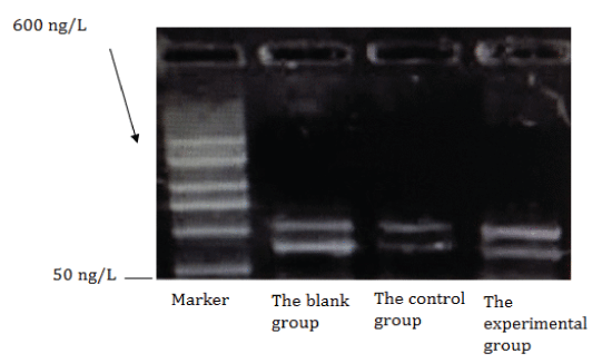
| The influence on the MCP-1 content on rats with microcirculatory disturbance Experimental results show Rhizoma zedoariae group is better than the nitroglycerin group (P<0.05), and the nitroglycerin group has the same effect with the blank group (P>0.05) (Table 4 and Figure 1). | ||||||||
Discussions |
||||||||
| MAV is one of the common diseases in modern clinics, and it is also a common disease affecting the life quality. For many years, researchers have generally believed that the cardiac X syndrome may be associated with myocardial ischemia, pain hypersensitivity, female hormone deficiency, and vagal dysfunction caused by microvascular abnormalities. [6-12]. But at present it is believed MAV has two possibilities: one is (the larger blood vessels) unstable atherosclerotic plaque rupture, rupture and shedding inside epicardial coronary artery, and large amounts of lipid rich plaque debris and fibrins and aggregated platelet enter coronary microcirculation to form micro thrombus embolism [13-15]; and the second is the insitu thrombus inside the coronary micro vascular thrombosis of the deep myocardium (endocardials) [17]. Many studies have indicated that the inflammatory response is involved in the development, progression and deterioration of coronary heart disease, [16]. After the inflammatory response, oxidative stress results in the decrease of NO activity and the synthesis decrease [18], while the antioxidant vitamin C can improve the NO biological activity and improve the coronary flow reserve (CFR), and also confirms this mechanism [19]. Inflammation responses can make the thromboxane A2, monocyte chemotaxis protein 1 biological activity become elevated [20], which leads to the increase of platelet aggregation activity, so that the coronary resistance gets increased, and CFR function declines. Therefore, NO, angiotensin II, thromboxane A2, monocyte chemotactic protein 1 can all change CFR. | ||||||||
| According to "Fuxie inside, against the flesh interstices, blood stasis to forge drugs" put forward by the Chinese medicine master Ren Jixue professor, in the important pathogenic role application among the heart disease syndrome differentiation system, the advantage of traditional Chinese medicine’s overall concept and the differentiation theory gradually forms the treatment model centered by the “phlegm and blood stasis Fu evil" theory and provides a new diagnostic method for the early intervention, vascular events [24]. In the aspect of Chinese medicine research, according to many years of clinical observation, we take the "phlegm and blood stasis" as the theoretical basis, and consider that the phlegm and blood stasis lies in collaterals and propose the phlegm and blood stasis go throughout the MAV always, only due to the deficiency of vital qi, phlegm and blood stasis Fu evil cannot reach outside, forming symptoms of chest pain. Under the deficiency of vital qi, when phlegm and blood stasis V evil can go out, due to the mood, fatigue, cold and other external inductions, it shows symptoms of chest pain. Therefore, in the process of the whole MAV disease, to eliminate phlegm and blood stasis is very important, which not only removes the phlegm and blood stasis superficially, but also through oral drugs for a long time intervenes the phlegm and blood stasis". So our department based on the theory self produces the Rhizoma zedoariae to eliminate the phlegm and blood stasis Fu evil to make the microvascular angina fundamentally become effectively treated. The composition of Rhizoma zedoariae has curcuma zedoary, raw oysters, San-Qi, fried Atractylodes, astragalus, licorice, wherein the rhizoma gets rid of Fu evil mainly and raw oysters get rid of blood stasis mainly; while San-Qi and Atractylodes assist dissipating phlegm and removing blood stasis secondarily, strengthening drug efficacy and the licorice reconciles the various drugs as the medium. Because of Rhizoma’s strong phlegm-killing effect, long-term oral usage may hurt the vital qi, so with Huangqi not injuring the vital qi, thus suitable for long-term usage. | ||||||||
| The experimental study shows that in the aspect of nitric oxide increases, Rhizoma zedoariae group is better than the nitroglycerin group (P<0.05), and the nitroglycerin group is better than the blank group (P<0.05), so the dilation of coronary vessels can the improve coronary blood flow reserve. In the aspect of angiotensin II reduction: zedoary Rhizoma zedoariae group is better than the nitroglycerin group and the blank group (P<0.05), and the nitroglycerin group has the same therapy effect with the blank group (P>0.05). In the aspect of the thromboxane A2 reduction: Rhizoma zedoariae group is better than that of nitroglycerin group and blank control group (P<0.05) and the nitroglycerin group has the same therapy effect with the blank group (P>0.05), changing the coronary blood flow due to the reduction blood-vessel shrinking substances. In the aspect of MCP-1 reduction: Rhizoma zedoariae group is better than the nitroglycerin group and the blank group (P<0.05), and the nitroglycerin group has the same therapy effect with the blank group (P>0.05). The reduction of these two factors can lead to the decrease in the platelet aggregation activity, thereby reducing the coronary resistance to improve the CFR function. In summary, zedoary turmeric capsule through increasing nitric oxide content and reducing vascular nerve agent II, reducing thromboxane A2 and mononuclear-cellized protein-1 and through a variety of mechanisms, improves coronary blood supply and improves the coronary flow reserve so as to treat the microvascular angina. But this experiment only in the inflammatory factor gives a preliminary study of its efficacy, and looks forward to the support from the further clinical verification data. | ||||||||
Tables at a glance |
||||||||
|
||||||||
Figures at a glance |
||||||||
|
||||||||
References |
||||||||
|
