ISSN: 0970-938X (Print) | 0976-1683 (Electronic)
Biomedical Research
An International Journal of Medical Sciences
Research Article - Biomedical Research (2017) Volume 28, Issue 12
The value of multi-slice spiral CT portography combined with functional magnetic resonance imaging observation of brain activity on the degree of liver cirrhosis and the prediction of hepatic encephalopathy risk
Xue Feng Jiang1, Lei Ding2* and Yuan Tian3
1Department of Gastroenterology, China-Japan Union Hospital of Jilin University, Xiantai Street No.126, PR China
2Department of Radiology, China-Japan Union Hospital of Jilin University, Xiantai Street No.126, PR China
3Departement of Medical Examination, China-Japan Union Hospital of Jilin University, Xiantai Street No.126, PR China
- *Corresponding Author:
- Lei-Ding
Department of Radiology
China-Japan Union Hospital of Jilin University
PR China
Accepted date: April 24, 2017
Objective: We sought to analyse the value of the multi-slice spiral CT portography combined with functional Magnetic Resonance (MR) imaging observation of brain activity on the degree of liver cirrhosis and the occurrence risk of Hepatic Encephalopathy (HE).
Methods: 68 cases of liver cirrhosis selected from our hospital during March, 2011-March, 2014 were divided into mild HE group (n=26), severe HE group (n=19) and non HE group (n=23) according to the degree of HE while another 30 cases of healthy individuals were also chosen as normal control group. All the subjects accepted multi- slice spiral CT portography combined with functional MR examination. Compared angiographic results with brain activity test results to analyse its value on the degree of liver cirrhosis indication and HE prediction.
Results: The multi-slice spiral CT portography, which has a good display effect on liver cirrhosis, can clearly indicate the intravenous course and the extent of disease; compared with the normal control group, patients were with significantly thicker vein diameter (p<0.05), while there were no significant differences in vein diameter when compared between patient groups (p>0.05); CT portography results classification: 28 cases for grade one, 30 cases for grade two and 10 cases for grade 3, no significant differences compared with Child-pugh grade (p<0.05); there were differences for the ALFF results in several brain regions between groups.
Conclusion: The multi-slice spiral CT portography can clearly show liver morphology and changes in collateral circulation. It also helps determine the severity of cirrhosis. The functional Magnetic Resonance (MR) imaging is helpful to identify the abnormal state of neuronal activity in brain regions and to judge the degree of HE and the risk of getting this disease.
Keywords
Multi-slice spiral CT, Portal vein angiography, Magnetic resonance imaging, Brain activity, Liver cirrhosis, Hepatic encephalopathy, Prediction
Introduction
Viral hepatitis, cholestasis, alcoholism can lead to pathological changes of liver tissues like diffuse fibrosis, pseudolobule occurrence, regenerative nodule formation, which are main causes of liver cirrhosis [1]. Hepatic Encephalopathy (HE), which could eventually lead to altered consciousness, coma or even death after causing patients brain dysfunction on the basis of metabolic and neurological disorders, is one of the most common and the most serious complications at the advanced stage of liver cirrhosis [2]. Therefore, the observation of the degree of liver cirrhosis and the prediction of HE risk is an important part of guiding clinical treatment programs and early prognosis. However, current clinical evaluation on patients is mainly relying on Child-pugh classification and other biochemical markers, which couldn't have ideal prognostic evaluation effects [3,4]. Thus, we did a clinical research by combining multi-slice spiral CT portography with functional Magnetic Resonance (MR) imaging.
Materials and Methods
Clinical materials and subjects
We selected 68 cases of liver cirrhosis patients in our hospital from March 2011 to March 2014, aging from 18 to 70 and patients while patients with central nerve system disorders, serious heart, lung, kidney and other important organ diseases, serious infection or malignancy and pregnant or lactating women were excluded. Among all the qualified subjects, 37 were male, 31 were female, age from 19-68 with an average age of 51.8 ± 7.2. We selected another 30 cases of healthy individuals with 15 males and 15 females, aging from 18-70 with an average age of 52.9 ± 10.6 to join the normal control group after signing a consent form. There were no statistical differences between general clinical data like age and gender of subjects (p>0.05).
This study was approved by the Ethical Clearance Committee of China-Japan Union Hospital of Jilin University. There were no conflicts of interest.
Examination
Grouping: The degree of HE for subjects in patients’ groups were determined by examining their blood ammonia, EEG, psychological intelligence, and EEG evoked potentials [5]. Then we divided the subjects into the mild HE group (n=26), severe HE group (n=19) and the normal control group (n=23) according to the results.
Multi-slice spiral CT portography: Portography was conducted by 320-slice spiral CT equipment (Toshiba, Japan). Contrast agent was Iohexol. The dose of contrast agent is 300 mg/ml and the rate of injection is 3.5-4.0 ml/s; scanning parameters were 120 kv, 280-370 mA, with a slice thickness of 5 mm and spiral distance of 0.516-1.375:1; scanning time for hepatic artery phase, portal venous phase and equilibrium phase were 25-30 s, 45-60 s and 120 s after contrast agent injection respectively; three-dimensional blood vessels were rebuilt by Maximum Intensity Projection (MIP), Volume Rendering (VR), Multi-Planar Reconstruction (MPR) methods after reconstructing images of a thickness of 0.625 mm [6]; final images were interpreted by two specialized radiologists in our hospital and they got a comprehensive determination by double-blind method and a third radiologist was prepared for different ideas.
Functional magnetic resonance: Functional magnetic resonance was operated by Achieva/Intera 3.0T MR (Philips, the Netherlands). Subjects were told to lie calmly on the MR equipment with their head fixed on the 16 channel sensitive coils for a sequence scanning. Axial scanning: T1W, T2W, T2 FLAIR sequences; Sagittal position: T1W; scanning parameter: TR 7.5 ms, TE 3.7 ms, the inversion angel was 8°, the visual field was 230 × 230 mm with 150 layers and the thickness of scanning layer was 0; We got the functional images by GRE-EPI methods. Relative parameters were as follows: TR 2000 ms, TE 35 ms, inversion angel was 90°, visual field was 230 × 230 mm with 34 layers and 4 mm for each layer, scanning time was 8 min. The functional MR data were sorted and time sequences were converted into spectrum data by pre-treatment, data format conversion, correction, registration. We also calculated the ratio of original functional MR and the whole brain average functional MR [7-9] to get the standard ALFF result and by this result, we got the differences of original low frequency amplitude and whole brain low frequency amplitude.
Results analysis
CT results analysis: Live cirrhosis grading system was made by referring to the multi-slice spiral CT portography results [9]. Grade one: level 4-5 by portal development; grade two: level 3-4 by portal development and with 2 or less vein open; grade three: level 2-3 by portal development and with 2 or more vein open. We compared the differences of portal vein diameter of CT results between patients’ groups and normal control group and analysed the consistency of CT results and the child-pugh grade of patients groups.
Functional magnetic resonance imaging results analysis: We compared the brain regions existing differences and the significance of differences of the MRI results between subjects of mild group and severe group, mild group and normal control group.
Statistical analysis
Statistical analysis was conducted by SPSS 13.0, count data were analysed by t-test and the test standard was set to α=0.05. There is a significant difference when p<0.05.
Results
Multi-slice spiral CT portography results
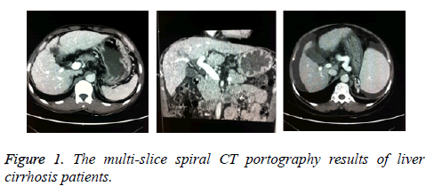
The multi-slice spiral CT portography has a good display effect of liver cirrhosis. The intravenous course and the extent of disease can be clearly shown through it (Figure 1).
Portal vein diameter
Compared with the normal control group, the portal vein diameter was significantly thicker in patient groups (p<0.05), while there were no significant differences in vein diameter when compared between patients’ groups (p>0.05, Table 1).
| Group | n | Portal vein diameter (cm) | p |
|---|---|---|---|
| Normal control | 30 | 1.195 ± 0.832 | <0.05 |
| Non HE | 26 | 1.538 ± 0.779a | |
| Mild HE | 19 | 1.655 ± 0.764a | |
| Severe HE | 23 | 1.701 ± 0.815a | |
| Note: acompared with the normal control group, p<0.05 | |||
Table 1: Portal vein diameter of all subjects (͞x ± s).
Multi-slice spiral CT portography grading and childpugh grading of liver cirrhosis patients
The multi-slice spiral CT portography grading result: 28 cases for grade one, 30 cases for grade two, 10 cases for grade three, no significant differences to child-pugh grading results (p>0.05, Table 2).
| Child-pugh grading | n | Multi-slice spiral CT portography grading | p |
|---|---|---|---|
| I | 31 | 28 | >0.05 |
| II | 26 | 30 | |
| III | 11 | 10 |
Table 2: Comparison of multi-slice spiral CT portography grading results and child-pugh grading results (n).
Functional MR results
There were differences in ALFF results in brain regions of right anterior medial frontal gyrus, left side of insula cover inferior frontal gyrus, right precuneus, right inferior temporal gyrus, left middle occipital gyrus, posterior lobe of right cerebellar hemisphere, anterior lobe of left cerebellar hemisphere between all groups. Check Tables 3 and 4 to see differences in functional MR results between mild HE group, severe HE group and normal control group.
| Brain region | Brodmann area | Voxel size (n) | MNI*coordinate (x, y, z) | tmax | |
|---|---|---|---|---|---|
| ALFF results increase | Right temporal inferior gyrus | 20 | 30 | (53, -49, -25) | 3.386 |
| ALFF results decrease | Posterior lobe of right cerebellar hemisphere | -- | 28 | (11, -89, -32) | -4.517 |
| Left side of insula cover inferior frontal gyrus | 44 | 37 | (-43, 8, 22) | -4.408 | |
| Right anterior medial frontal gyrus | 8 | 27 | (4, 22, 59) | -3.229 | |
| Note: *Institute of neuroscience of Montreal | |||||
Table 3: Checking results of brain regions existing differences in functional MR results between mild HE group and severe HE group.
| Brain region | Brodmann area | Voxel size (n) | MNI*coordinate (x, y, z) | tmax | |
|---|---|---|---|---|---|
| ALFF results increase | Right temporal inferior gyrus | 20 | 31 | (53, -46, -25) | 3.369 |
| Anterior lobe of left cerebellar hemisphere | -- | 20 | (-22, -43, -37) | 3.225 | |
| ALFF results decrease | Posterior lobe of right cerebellar hemisphere | -- | 28 | (10, -89, -36) | -4.174 |
| Left side of insula cover inferior frontal gyrus | 44 | 41 | (-47, 13, 25) | -4.112 | |
| Right precuneus | 23 | 22 | (11, -64, 23) | -4.197 | |
| Right anterior medial frontal gyrus | 8 | 27 | (4, 38, 49) | -3.368 | |
| Left middle occipital gyrus | 17 | 25 | (-10, -98, 0) | -2.994 |
Table 4: Checking results of brain regions existing differences in functional ALFF results between mild HE group and normal control group.
Discussion
The formation of liver cirrhosis is a slow process experiencing a number of pathological changes, which include chronic inflammation, fibrosis, cirrhosis and portal hypertension, etc. With the aggravation of pathological changes, prognosis will also become worse. And accompanied with pathological changes, there were also significant changes in liver function and morphology. Therefore, there is significance for morphological quantitative detection in the degree of liver cirrhosis judgment [10].
The worsening of liver cirrhosis is always associated with metabolic disorders causing more neurotoxic substances, which may influence on the structure and energy of neurons and glial cells and this serves as the main cause of HE. Many researchers found that although indicators like ammonia and neuropsychological test scores have some significance in HE diagnosis, their effects on HE occurrence risk and prognosis were not so obvious [11,12].
Multi-slice spiral CT portography is quite effective in terms of vascular display technology with its high time, space, and density resolution. This technology could clearly observe vascular arranging and portal vein branches. With changes in liver tissues morphology intensify, pathological changes in portal system become more serious. CT portography can effectively show changes like portal vein thinning, distortion or occlusion in liver and its results grading can determine the degree of liver cirrhosis to a certain extent [13]. Our study found that there was no significant difference in CT portography results grading and Child-pugh grading, which indicated the consistency of these two grading methods. And in clinic, patients’ Child-pugh grading was always significantly improved after treatment whereas the reversal of their morphological abnormalities in liver was not evident [14]. Therefore, it is more reliable to use CT portography to grade the severity of liver cirrhosis. What's more, compared with Digital Subtraction Angiography (DSA), CT potography has several advantages such as low incidence of complications, high scanning speed, good image reconstruction, clear observation of adjacent structures [15], which were more helpful for assessing the severity of patients’ liver cirrhosis and selecting treatment programs.
However, with the increase of portal pressure levels and the gradual opening of collateral circulation, the risk of getting hepatic encephalopathy in liver cirrhosis patients have significantly increased. CT portography has no significant guiding value in the occurrence risk of HE. To solve this problem, we chose functional MR to check the differences in brain regions of liver cirrhosis patients and healthy people and found that there were many changes of several brain regions in ALFF checking results for HE patients, indicating that changes in ALFF results may have significance in diagnosing and predicting the HE occurrence risk. Compared with the normal control group, there were decreases in the ALFF results of right anterior medial frontal gyrus and left side of insula cover inferior frontal gyrus of mild HE group patients. Since these two brain regions were the center for brain cognition and memory functions, decreases in their ALFF results suggested prefrontal damage in patients brains, which may further cause low attention regulatory capacity, spatial working memory impairment, spontaneous neuronal activity weakened state in resting state as well as influencing cognitive competence of patients [16,17]. For severe HE patients, there were decreases in the functional ALFF results of precuneus and cuneus brain regions indicating that they had more severe neurological damage. Studies have shown that precuneus and cuneus brain injury can cause the default mode network (DMN) dysfunction [18], which in turn cause the brain function capability significantly to reduce while maintaining the resting state of the brain and the occurrence of brain endogenous functional organization damage. Besides, there were increases in the functional ALFF results of the right inferior temporal gyrus and the anterior lobe of left cerebellar hemisphere of severe HE patients. We considered that it was related to compensatory function of brain-related functional areas and its mechanism remains to be further explored.
In conclusion, multi-slice spiral CT portography can identify liver morphology and collateral circulation changes helping the determination of the degree of liver cirrhosis, while the examination of brain activity by functional MR is helpful to identify abnormal status of brain neuronal activities and is valuable to judge the degree and risk of HE occurrence.
References
- Montano-Loza AJ. New concepts in liver cirrhosis: clinical significance of sarcopenia in cirrhotic patients. Minerva Gastroenterol Dietol 2013; 59: 173-186.
- Romero-Gomez M, Montagnese S, Jalan R. Hepatic encephalopathy in patients with acute decompensation of cirrhosis and acute on chronic liver failure. J Hepatol 2014.
- Holecek M. Ammonia and amino acid profiles in liver cirrhosis: Effects of variables leading to hepatic encephalopathy. Nutrition 2014.
- Dhiman RK, Kurmi R, Thumburu KK, Venkataramarao SH, Agarwal R. Diagnosis and prognostic significance of minimal hepatic encephalopathy in patients with cirrhosis of liver. Dig Dis Sci 2010; 55: 2381-2390.
- Karsdal MA. Review article: the efficacy of biomarkers in chronic fibroproliferative diseases-early diagnosis and prognosis, with liver fibrosis as an exemplar. Aliment Pharmacol Ther 2014; 40: 233-249.
- Savale L, Sattler C, Sitbon O. Pulmonary hypertension in liver diseases. Presse Med 2014; 43: 970-980.
- Suzuki T, Shinjo S, Arai T, Kanai M, Goda N. Hypoxia and fatty liver. World J Gastroenterol 2014; 20: 15087-15097.
- Lemoinne S, Thabut D, Housset C, Moreau R, Valla D. The emerging roles of microvesicles in liver diseases. Nat Rev Gastroenterol Hepatol 2014; 11: 350-361.
- Hope TA, Ohliger MA, Qayyum A. MR imaging of diffuse liver disease: from technique to diagnosis. Radiol Clin North Am 2014; 52: 709-724.
- Bowlus CL, Gershwin ME. The diagnosis of primary biliary cirrhosis. Autoimmun Rev 2014; 13: 441-444.
- Jaafar J, de Kalbermatten B, Philippe J, Scheen A, Jornayvaz FR. Chronic liver diseases and diabetes. Rev Med Suisse 2014; 10: 1254, 1256-1260.
- Kobelska-Dubiel N, Klincewicz B, Cichy W. Liver disease in cystic fibrosis. Prz Gastroenterol 2014; 9: 136-141.
- Watson H. Satavaptan treatment for ascites in patients with cirrhosis: a meta-analysis of effect on hepatic encephalopathy development. Metab Brain Dis 2013; 28: 301-305.
- Dhiman RK. Gut microbiota and hepatic encephalopathy. Metab Brain Dis 2013; 28: 321-326.
- Hou D. Multi-slice computed tomography 5-minute delayed scan is superior to immediate scan after contrast media application in characterization of intracranial tuberculosis. Med Sci Monit 2014; 20: 1556-1562.
- Chen X. Clinical value of multi-slice spiral computed tomography angiography and three-dimensional reconstruction in the diagnosis of double aortic arch. Exp Ther Med 2014; 8: 623-627.
- Liu WN. The value of multi-slice spiral computed tomography portography in assessing severity of liver cirrhosis and predicting episode risks of hepatic encephalopathy. Zhonghua Gan Zang Bing Za Zhi 2014; 22: 509-513.
- Sheasgreen C, Lu L, Patel A. Pathophysiology, diagnosis, and management of hepatic encephalopathy. Inflammopharmacology 2014; 22: 319-326.
