ISSN: 0970-938X (Print) | 0976-1683 (Electronic)
Biomedical Research
An International Journal of Medical Sciences
- Biomedical Research (2011) Volume 22, Issue 2
Vascular Foramina of Scaphoid and its Clinical Implications
P.P Dubey*, Navneet Kumar Chauhan, M.S. Siddiqui
Department of Anatomy , Chattrapati Shahuji Maharaj Medical University U.P., Lucknow. (U. P.)
Department of Anatomy, Era Medical Collage, Lucknow (U.P.), India
Accepted date: December 13 2010
Keywords
Scaphoid, Foramen, Carpal, Wrist, blood supply
Introduction
The blood supply of carpal bone has been rich field for the workers in the 19th century. The vascular pattern put forward in correlation to gross anatomy, radiographic pat-tern of extraosseous and intraosseous blood vessels, ob-serving the vascular foramina on the bones has been de-scribed. Obletz & Halbstein [1] studied the vascular fo-ramina visible on dried specimens of the human scaphoid and proposed a widely accepted concept of a predomi-nantly distally based blood supply entering through the dorsal ridge. However, this concept has not been entirely supported by the results of more recent studies carried out by various investigators. The dorsal, laterovolar and dis-tal group of feeding vessels enter in respective side of scaphoid bone [2] .The vasculature perforate the dorsal area of the scaphoid bone through numerous small foram-ina along the spiral groove and dorsal ridge [3].
The fracture and fraction dislocation of scaphoid is very common in wrist trauma and avascular necrosis is directly connected with vascular pattern of scaphoid.[4] In two-thirds of all cases, the scaphoid bone is most frequently fractured bone of the wrist, and it is uniformly vascular-ized, so that by fracture both fragments retain their blood supply. In the remaining third of all cases, only one end of the scaphoid is supplied arterially. Thus the poorly vascu-larized fragment frequently becomes necrotic [5]
We decided to study the vascular foramina as a parameter of blood supply of scaphoid and its possible relationship to fracture healing.
Material and Methods
The vascularization of scapoid bone was estimated by counting of vascular foramina on dry scaphoid bone of adult age group of both sexes. The dry scaphoid bones were obtained from Department of Anatomy, Moti Lal Nehru Medical College, Allahabad (U.P.), Era Medical College,Lucknow(U.P.) and Chattrapati Shahuji Maharaj Medical University U.P. Lucknow. A total number of 220 scaphoid bones of both sides were selected for study. The non-articular along with articular surfaces of scaphoid bone were considered for observations. The foramina of scaphoid were taken as site of vessel entry. These foram-ina were counted on the surface of bones with the help of operating loop lens.
Observations
Dry, 220, scaphoid bones of both sides were taken for observations in present study. The scaphoid bones were not differentiated according to sex. The vascular foramina were found only on palmar and dorsal surfaces of sca-phoid. The articular surfaces were devoid of any vascular entry. According to distribution of foramina, observations were grouped as under:
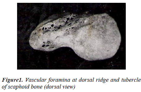
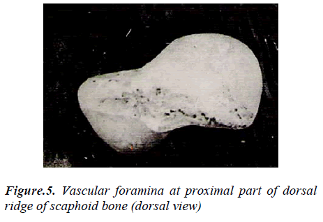
1. The numbers of foramina on the dorsal surface were counted on scaphoid bone. It was observed that these foramina are ranged from 1to10 in 14 (6.36%) specimens; foramina ranging 11to20 were present in 144 (65.45%) specimens and 21to32 fo-ramina in 62 (28.18%) specimens. (Table 1; Figures 1,5)
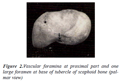
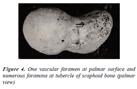
2. The number of vascular foramina on dorsal surface of scaphoid ranged between 4-32 whereas on pal mar surface 1- 8 (Table 4) (Figures 2,4).
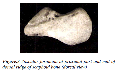
3. It was observed that 6% specimens show no vascu-lar foramina proximal to the mid of the waist while 32.72% specimens show one vascular foramen proximal to the mid of waist and 61.28% (Figure-2,3) specimens show more than one vascular foramina proximal to the mid of waist (Figure 3). Number of vascular foramina 1 to 2 distal to mid of waist 17.72% was present in specimens, and more than two in 82.28% specimens. (Tables 2,3)
Discussion
In present study vascular foramina are noted in distal and proximal part on both palmar surface and dorsal surface of scaphoid. The vascular foramina distal to mid of waist are 1 to 2 in 17.72 % specimen and rest of bones having more then 2 foramina are 82.28%.There is no scaphoid without vascular foramina distal to mid of waist. But the proximal part to mid of waist of scaphoid are without vascular foramina in 6% cases, one vascular foramina in 32.72% and more than one in 61.28% cases. These foramina clearly suggest that the proximal part of scaphoid receives less blood supply. It can be predicted that 6% scaphoid are highly vulnerable and 32.72% scaphoid are prone to avascular necrosis according our finding. Obletz & Halbstein (1938) described that 67% of individuals have 2 or more vascular foramina proximal to the waist, 20% have only one foramen proximally and 13% were found to have no proximal foramina. Thus, in one third of waist (mid) fractures, there will be dimin-ished blood supply to the proximal fracture fragment this may produce a non-union and avascular necrosis may ensue.[1]. In present study 6% scaphoid were found with-out vascular foramina on proximal part. The blood supply of the scaphoid comes from radial artery. The proximal pole and 70-80% of bone receives their blood supply from vessels entering through dorsal ridge. The tuberosity and distal 20-30% of the bone are supplied by volar branches of radial artery and its superficial palmar branch [6] .The dorsal, proximal, near base of tubercle and radial part of volar surface of scaphoid is penetrated by branches of radial arteries .The numerous small vessels enter in sca-phoid through the radio volar aspect [7]. These findings are similar to our finding; however Barber(1972) noted three groups of vessels, entering at various intervals along the dorsal ridge and branching to supply the tuberosity and the proximal pole [8]. He did not find any vessels entering the scaphoid through its volar surface. According to Fasal et al(1978) a branch of the arteria radialis or of the ramus dorsalis arteriae radialis enters the dorsal sur-face in the middle and distal third by several foramina whereas the proximal third of the scaphoid showed no vascular foramen [9].The location and distribution of the vascular foramina visible along the dorsal ridge of a dried scaphoid would account for the occurrence and incidence of aseptic necrosis of the proximal pole of the scaphoid following fracture through the waist.[10] Our series con-sistently demonstrated the presence of foramina confined to the dorsal ridge of the scaphoid. These did not corre-spond to the division into a dorsal and laterovolar arterial supply of the proximal pole suggested by Taleisnik & Kelly (1966) [2] .The main blood vessels came from the dorsal and the radial side of wrist and passed through the whole scaphoid along the crest of scaphoid.[11] .A good blood supply of the scaphoid bone from palmar, dorsal and radial vessel groups with a variety of anastomoses provide sufficient collateral blood flow from adjacent regions . Since blood supply is available from the palmar circulation, a dorsal approach to the scaphoid bone is pos-sible. [12]. Osteonecrosis is said to occur in 13%–50% of cases of fracture of the scaphoid, and the incidence of osteonecrosis is even higher in those with involvement of the proximal one-fifth of the scaphoid. [4]. Dawson et.al.(2001) found that the rate of nonunion of scaphoid fracture is 12% which is similar to other studies (10% to 13%) when fractures of the scaphoid were treated conser-vatively. [13].
Finally we can conclude that 6% scaphoid are without foramina, 32.72% scaphoid with one foramen and 61.28% scaphoid with more than one foramen on proximal part.
The dorsal surface has four times more foramina than palmar surface .These observations indicate that there is higher blood supply in proximal part of scaphoid in com-parison of other studies. The surgical approach for sca-phoid may be from both dorsal and palmar aspect as vas-cular foramina present in both surface of scaphoid.
References
- Obletz BE, HaIbstein BM. Non-union of fractures of the carpal navicular. J Bone Joint Surg; 1938; 20:424-8.
- Taleisnik.J, Kelly P. The extraosseous and intraosse-ous blood supply of the scaphoid bone. Journal of Bone and Joint Surgery; 1966; 44(A), 1125-1137
- Gelberman, PH, Menon J. The vascularity of the sca-phoid bone. J Hand Surg 1980; 5: 508-513
- Scott P. Steinmann and Julie E. Adams Scaphoid frac-tures and nonunions: diagnosis and treatment J Orthop Sci. 2006; July; 11(4): 424-431
- Oehmke HJ. Blood supply and function of the sca-phoid bone Unfallchirurgie. 1987 Jun; 13(3): 174-177.
- Gelberman RH, Bauman TD, Menon J. The vascular-ity of the Lunate bone and Kinebock’s Disease. J Hand Surg 1980; 5: 272-278.
- Gretive, s. Arterial anatomy of the carpal bones; Acta Anatomica.1955; 25: 331-345.
- Barber, H.The intraosseous arterial anatomy of the adult human; Carpus orthopaedics.1972; 5: 1-19.
- Fasol P, Munk P,StricknerM. Blood supply of the scaphoid bone of the hand; Acta Anat (Basel) 1978; 100(1): 27-33
- Handley RC, Pooley J. The venous anatomy of sca-phoid. J.Anat.1991; 178: 155 -118.
- Kong WY, Xu YQ, Wang YF, Chen SC, Liu ZL, Li XG; Anatomic measurement of wrist scaphoid and its clinical significance Chin. J. Traumatol. 2009; 12(1): 41-44.
- Oehmke MJ, Podranski T, Klaus R, Knolle E, Wein-del S, Rein S, HJ. The blood supply of the scaphoid bone. J Hand Surg Eur 2009; 34(3):351-357
- Dawson JS,. Martel AL, Davis TRC. Scaphoid blood flow and acute fracture healing;A dynamic MRI study with enhancemen with Gadolium; J Bone Joint Surg [Br] 2001; 83-B: 809-14

